Clinical:
- A 53 years old lady
- History of trauma one month ago
- Presented with left eye proptosis, redness and blurred vision


CT scan findings:
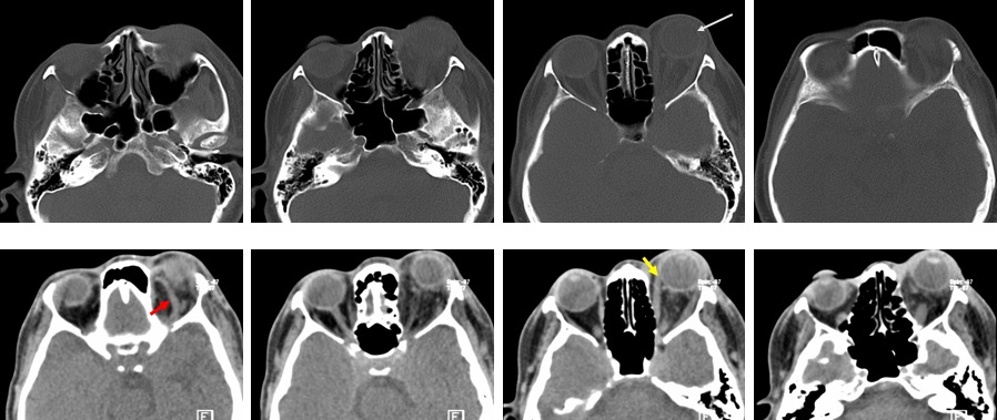
- There is left proptosis (white arrow).
- The left superior opthalmic vein is dilated (red arrow).
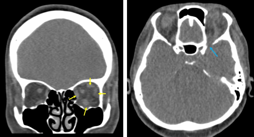
- There is enlargement of extra ocular muscles on the left side (yellow arrows).
- There is associated streakiness of intraconal fat.
- The left superior orbital fissure is also widened (blue arrow)

CTA findings:
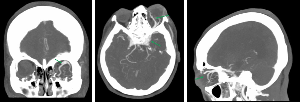
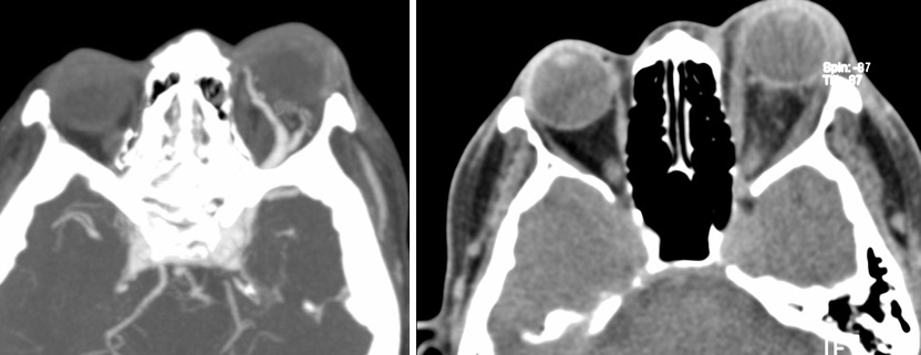
- Abnormal dilatation and opacification of left superior orbital vein
- There is abnormal bulging and opacification of cavernous sinuses; worse on the left side
- No abnormal enlargement of cortical veins
Diagnosis: Traumatic carotid cavernous fistula
Discussion:
- Carotid cavernous fistula is an abnormal communication between internal carotid artery and veins of cavernous sinus.
- Post traumatic, it can occur with laceration of ICA within cavernous sinus usually secondary to basal skull fracture (not demonstrated in this patient).
- Presentation include pulsating exophthalmos, chemosis, conjunctival edema, persistent orbital bruit, restricted extra ocular movement and decreased vision.
- CT findings include enlarged oedematous extraocular muscles, dilatation of the superior ophthalmic veins, facial veins or internal jugular veins with focal or diffuse enlargement of cavernous sinus.
- Occasionally sellar erosion or enlargement can also be seen.
- Enlargement of superior orbital fissure is seen in chronic cases.
- Angiography shows ipsilateral ICA contrast injection shows wall of ICA to be incomplete, contralateral ICA contrast opacification and compression of involved ICA, early opacification of veins of cavernous sinus and retrograde flow through dilated superior ophthalmic vein.

Recent Comments