
Case contribution: Dr Radhiana Binti Hassan
Clinical:
- A 33 years old female
- Day 19 post LSCS, presented with abdominal pain
- No hematuria, no fever
- Ultrasound shows no abdominal collection
- Incidental finding of right renal calculi
CT scan findings:
- Acalculus cholecystitis as the cause of current presentation
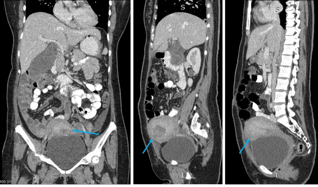
- Other findings: Post operative uterus (blue arrows)
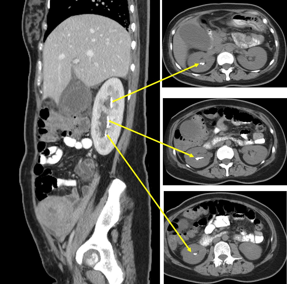
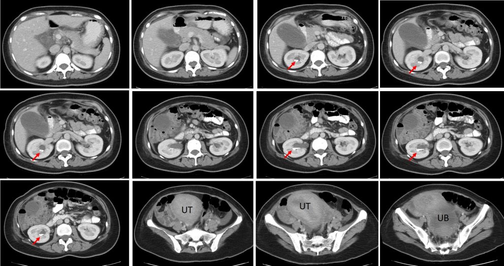
- There are multiple hyperdense foci in the pelvicalyceal system of right kidney (arrows) involving the upper, lower and interpolar.
- These hyperdense foci are seen layered at its dependant part.
- A small calculus is also seen in the left lower pole. Otherwise there is no hydronephrosis bilaterally.
- No ureteric calculus is seen.
- Urinary bladder is well distended.
Diagnosis: renal milk of calcium
Discussion:
- Milk of calcium is a viscous colloidal suspension of calcium carbonate, calcium phosphate, calcium oxalate and occasionally ammonium phosphate.
- These are seen in patients with calyceal diverticulum or in patients with multiple radiodense levels of milk of calcium in hydronephrotic kidney.
- The etiology is uncertain but obstruction and inflammation seems to be the key factors.
- Obstruction and stagnation of urine possibly result in super saturation of calcium salts resulting in the formation of calcium microliths. Due to a disturbance in stone forming and inhibiting factors a dynamic equilibrium probably results, preventing the aggregation of the microliths. Why the microliths do not increase in size and form a stone remains unexplained.
- The importance of MOC recognition is to avoid unnecessary procedures like ESWL or PCNLs or any other unwarranted interventions.
- A non-contrast CT scan is able to demonstrate the layers of calcified material in the dependant portion of the calyceal system with characteristic postural change on supine and prone positions.
Recent Comments