
Case contribution: Dr Radhiana Binti Hassan
Clinical:
- A 56-years old male
- Underlying lung carcinoma and hypertension
- Presented with painless and gradual blurred vision right eye
MRI orbit findings:
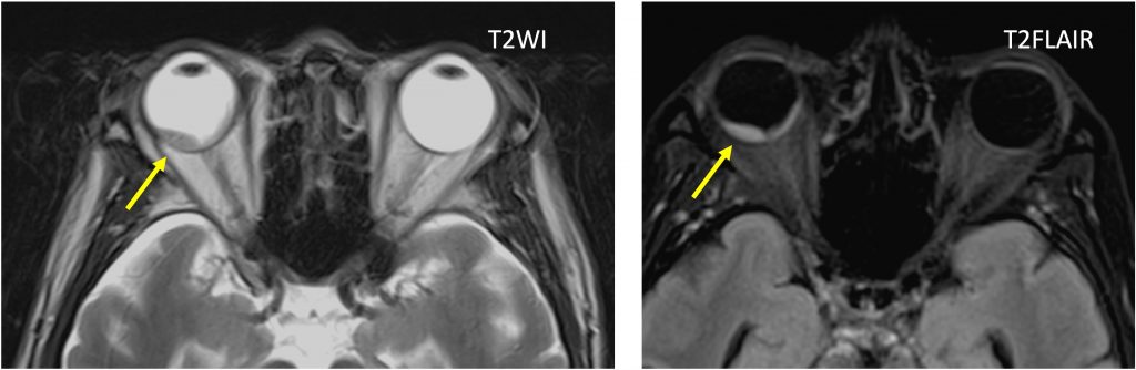
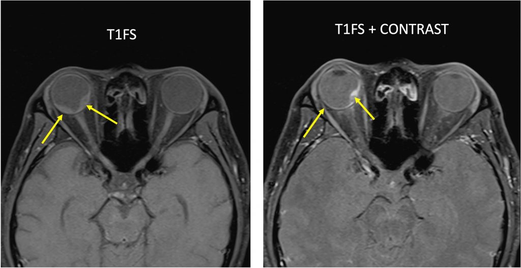
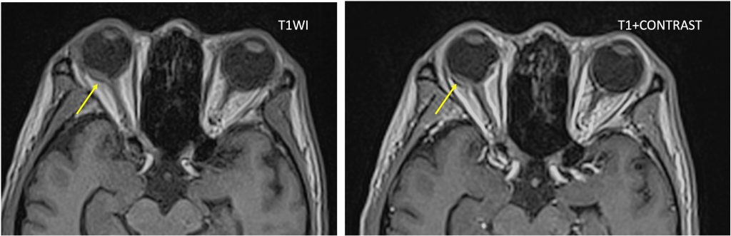
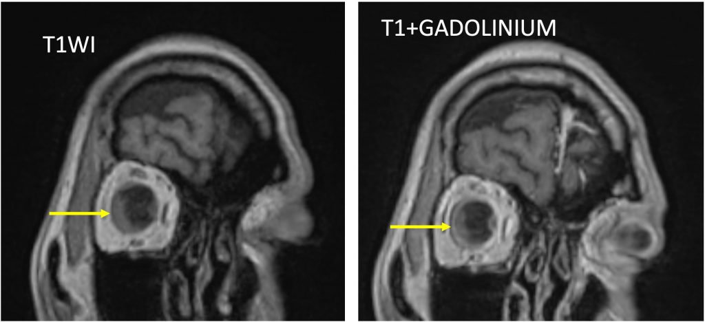
- There is mass lesion seen within the right globe (yellow arrow)
- The lesion is isointense to grey matter on T1WI, T2WI and T1fat-suppression sequence.
- It shows heterogenous enhancement post contrast.
- The lesion is mainly located at posterior part of the globe extending anteriorly along the retina with biconvex margin.
- Both globes maintains its normal shape and configuration.
- No involvement of the lens or anterior chamber region.
- The optic nerve and extraocular muscles are normal in appearance.
- No mass lesion within intraconal or extraconal space.
- Lacrimal glands are also normal.
- No proptosis.
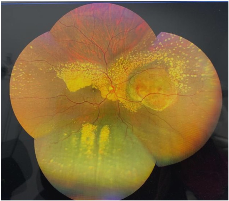
Funduscopy:
- suggestive of metastasis
Diagnosis: Ocular metastasis
Discussion:
- Ocular metastasis account for over 80% of ocular pathology
- It can be bilateral up to 25% of cases
- It is non-calcified masses within the orbit
- It is distinctly different from extraocular orbital metastasis
- It is also known as uveal metastasis
- The most common primary sites are breast carcinoma, lung carcinoma, GI carcinoma, cutaneous melanoma and neuroblastoma
Recent Comments