
Case contribution: Dr Radhiana Hassan
Clinical:
- A 30- years old female
- Underlying multiple medical conditions
- Pseudohypoparathyroidisms with hypocalcemia under endocrine htaa
- Vitamin d deficiency
- Psoriasis
- Presented with painful right thigh with limited movement due to pain
- Clinical examination shows reduced range of motion hip and knee due to pain
- Also had stiffness due to immobility
- Tender at anterior compartment . Lateral and medial compartment is normal. No tenderness at bony prominence
Radiographic findings:
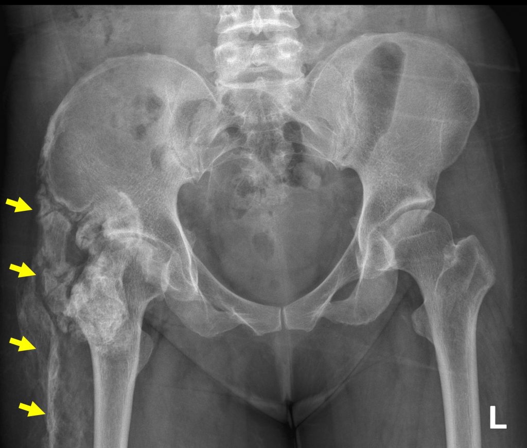
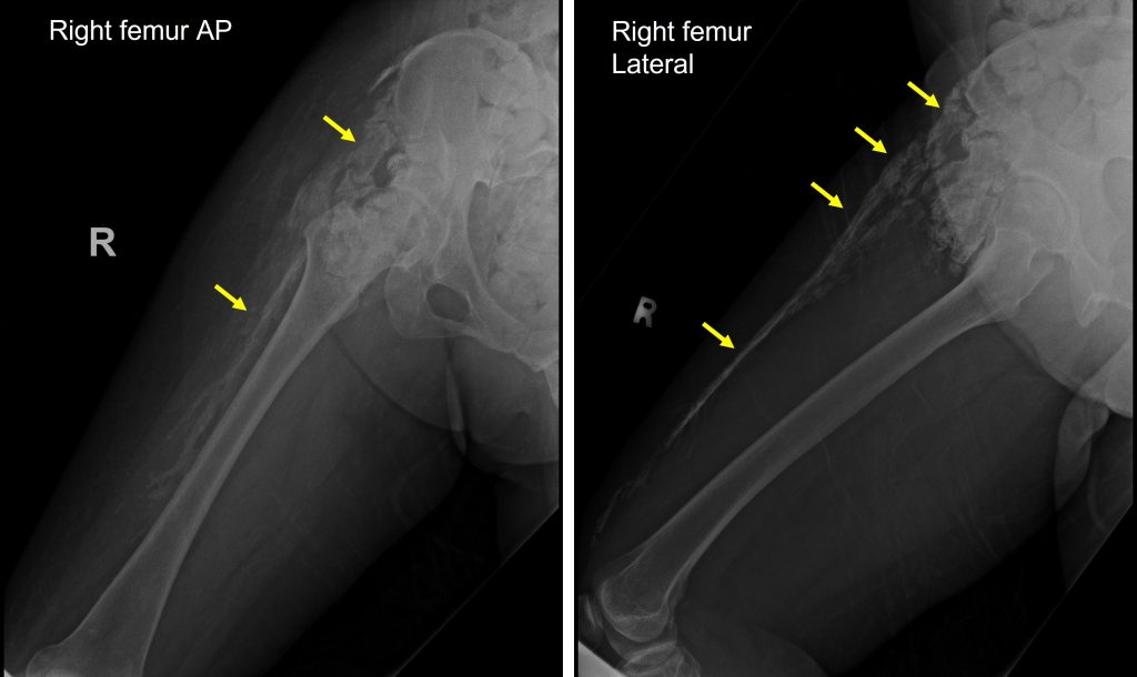
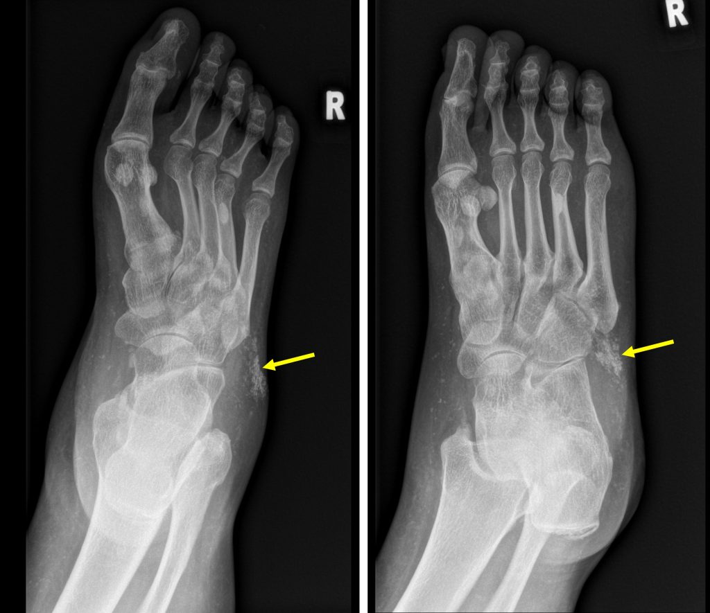
- Heterotopic calcifications are seen within the soft tissue
- At anterolateral region of right thigh and
- lateral foot and ankle region
- Associated mild heterogenous soft tissue swelling
Diagnosis: myositis ossificans
Discussion:
- It is the most common form of heterotopic ossification
- It usually occurs within large muscles
- It is important to recognize this condition which is considered as skeletal “don’t touch lesions” and differentiate it with aggressive pathological diseases.
- Some conditions related to this are:
- Myositis ossificans circumscripta- new bone formed usually after trauma
- Myositis ossificans progressive – a rare autosomal dominant disorder
- Panniculitis ossificans- similar to myositis ossificans but occur in subcutaneous tissue
- Fibro-osseous pseudotumour of the digits- variant of myositis ossificans in the fingers and toes
- Initially, on imaging can be seen as soft tissue swelling but no obvious calcification
- Calcification is apparent within 2-6 weeks
- Classic appearance of well circumscribed peripherally calcified appearance by 2 months
- It shows cleft between the adjacent bone but may be difficult to see on plain radiograph
- CT shows the mineralization proceeding from the outer margin towards center better.
- Differential diagnosis: parosteal osteosarcoma and soft tissue sarcoma
Recent Comments