
Clinical:
- A 46 years old man
- Involved in motor vehicle accident
- History of loss of consciousness
- Complains of headache and persitent vomiting
- Clinical assessment in ED shows GCS 15/15, no neurological deficit
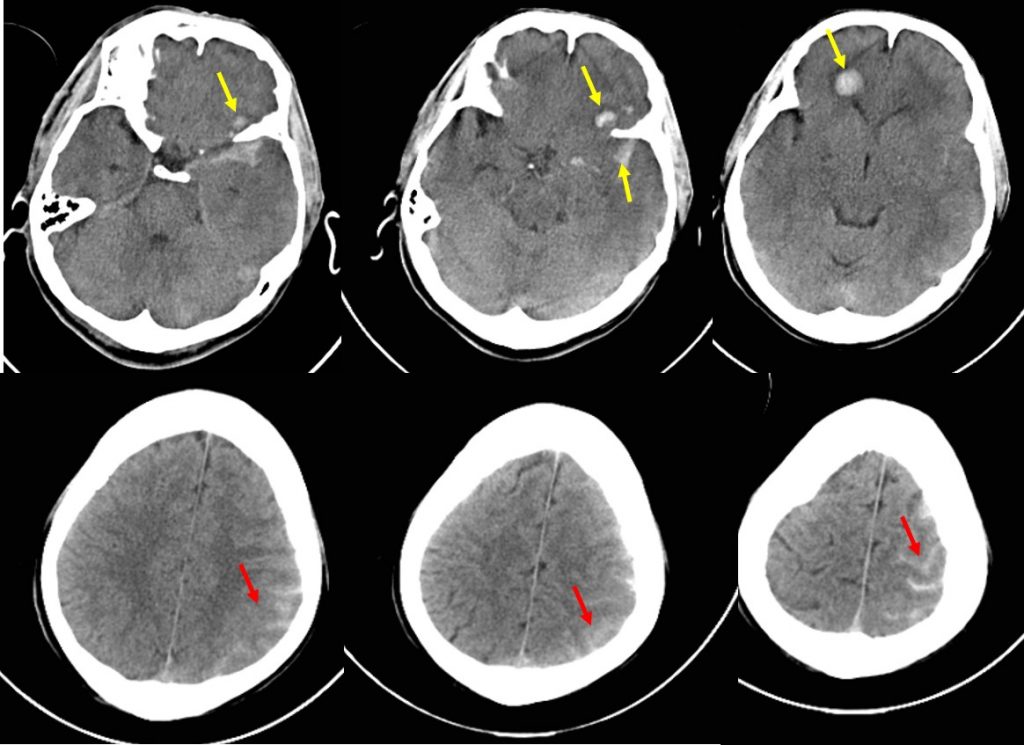
CT findings:
- There are multiple hyperdense foci at left frontal and temporal lobe (yellow arrows).
- The hyperdensities are of various sizes with minimal surrounding hypodensities.
- There are hyperdensities along the cerebral sulci at left frontoparietal region (red arrow)
- There is associated effacement of the cerebral sulci.
- No midline shift, no internal brain herniation
Diagnosis: Traumatic brain injuries (cerebral contusion and subarachnoid hemorrhage)
Discussion:
- Cerebral contusion also known as brain bruise occurs due to closed head injury against inner surfaces of the skull at the time of impact
- Usually at contrecoup site of the injury
- Location of cerebral contusion is at anterior base frontal, temporal lobes
- CT appearance show small, focal areas of petechial haemorrhage, peripherally located, multiple and can be bilateral. However CT scan can be normal, very subtle and/or odema on initial imaging. Over time, about half of the contusions evolve and grow larger in size.
- Traumatic subarachnoid hemorrhage (SAH) occurs in about 40% of patients with moderate to severe head injury is associated with a significant worse outcome
- Nearly all cases of traumatic SAH have other lesions to suggest traumatic cause
- CT findings of SAH is seen as linear areas of hyperdensities in the cerebral sulci and convexities, Sylvian fissures or basilar cisterns. Subtle SAH can be seen at interpeduncular fossa.
Recent Comments