Image 1

Questions:
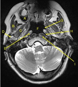
- Name the examination.
- Name the labelled structures.
Answers:
- MRI of neck in axial plane, T2-weighted image.
- The labelled structures:
- Right maxillary sinus
- Right masseter muscle
- Right lateral pterygoid muscle
- Right head of mandible
- Right torus tubarius
- Nasopharynx
- Left Eustachian tube
- Left lateral pharygeal recess (fossa of Rosenmuller)
- Prevertebral muscle (longus capitis muscle)
- Medulla oblongata
Image 2

Questions:
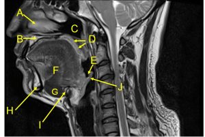
- Name the examination.
- Name the labelled structures.
Answers:
- MRI of neck, sagittal plane in T2-weighted image.
- The labelled structures:A. Nasal turbinateB. Hard palateC. Nasopharynx
D. Soft palate
E. Vallecula
F. Genioglossus muscle
G. Mylohyoid muscle
H. Mandible
I. Hyoid bone
J. Epiglottis

Recent Comments