Case contribution: Dr Radhiana Hassan
Clinical:
- An 18 years old boy with epilepsy
- Recent admission for status epilepticus
- Found unconscious at home, CPR by parents
- In ED GCS=E1V2M2, also had 2 episodes of GTC seizures
- Intubated and admitted to ICU
- Poor GCS recovery after 10 days in ICU

CT Findings:
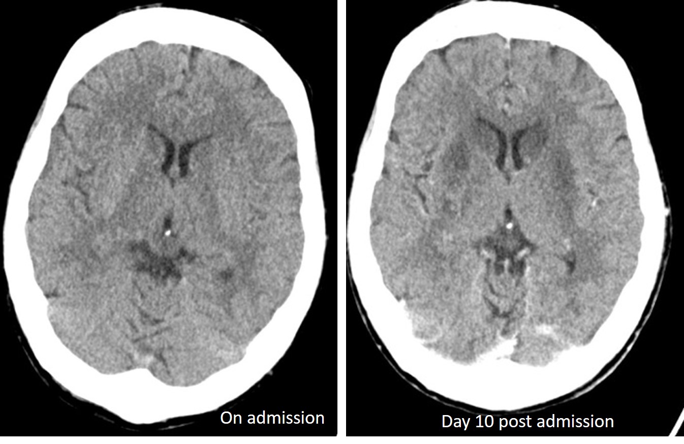
- CT scan of the brain shows bilateral almost symmetrical hypodensities involving the basal ganglia.
- The changes were not seen on previous CT scan done during admission.

MRI findings:
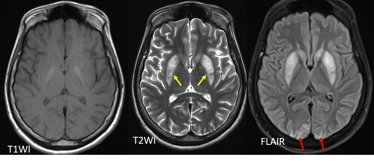
- MRI shows signal abnormalities in both basal ganglia (yellow arrows) as noted on CT scan.
- Subtle hyperintensities are also seen at cortical region of both occipital lobe (red arrows).
Diagnosis: Global hypoxic-ischaemic injury
Discussion:
- also known as adult hypoxic-ischaemic encephalopathy
- typically present following an acute event causing hypoxaemia or hypoperfusion such as near drowning, asphyxia, cardiac or respiratory arrest
- primarily affects the grey matter structures; basal ganglia, thalami, cerebral cortex, cerebellum and hippocampi
- As seen in this case, CT can be normal at early stage
- CT findings include diffuse edema with effacement of the CSF-containing spaces, decreased cortical gray matter attenuation with loss of normal gray-white differentiation, and decreased bilateral basal ganglia attenuation
- The reversal sign and the white cerebellum sign may be seen and indicate severe injury with a poor prognosis
- Diffusion-weighted MR imaging is the earliest imaging modality to become positive, usually within the first few hours after a hypoxic-ischemic event.
- T2-weighted images typically become positive during early subacute period (24 hours–2 weeks)
- Gray matter signal intensity abnormalities at conventional MR imaging may persist into the end of the 2nd week.
- In the chronic stage, T2-weighted images may demonstrate some residual hyperintensity in the basal ganglia, and T1-weighted images may show cortical necrosis
Recent Comments