Clinical:
- A 17 years old boy
- Alleged MVA
- On arrival in ED, GCS 15/15, vital signs are stable

CT scan findings:
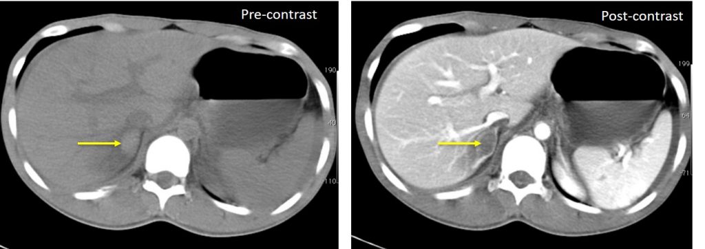
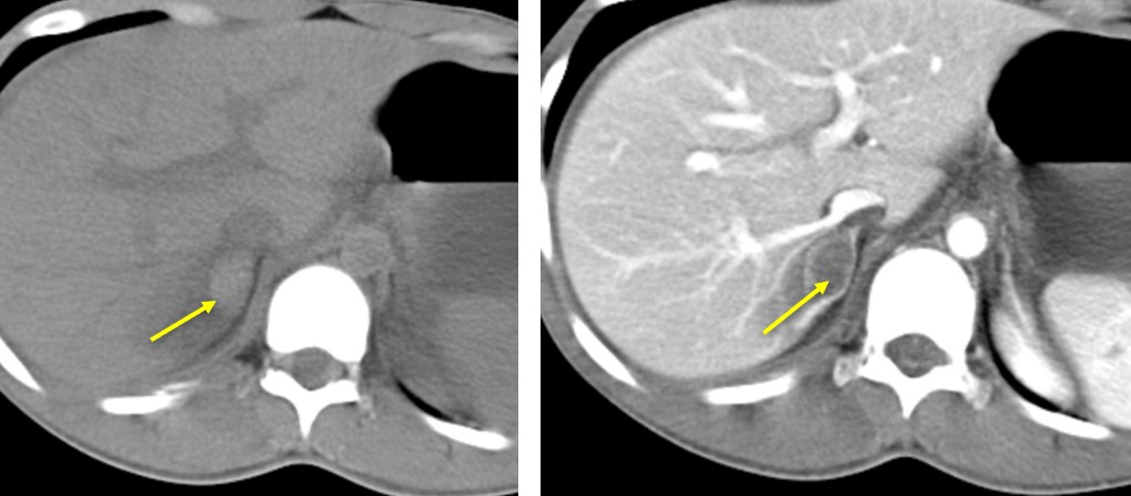
- An oval-shaped lesion at right adrenal region which is hyperdense on pre-contrast image
- Post contrast image shows the lesion to be relatively hypodense than the surrounding normal enhancement of right adrenal gland
- There is associated streakiness of the fat plane surrounding the right adrenal gland.
- This is in keeping with right adrenal hemorrhage.
Diagnosis: Traumatic right adrenal hemorrhage.
Discussion:
- Adrenal gland trauma is present on 1-2% of CT imaging in blunt trauma.
- Adrenal hemorrhage is the most common injury to the adrenal gland. Adrenal laceration is rare.
- Isolated adrenal trauma is uncommon, comprising <5% of cases.
- Associated injuries in adrenal gland trauma include lung injury, pneumothorax, solid abdominal organ injuries, ribs and spine fractures and head injuries.
- On ultrasound, an early-stage haematoma appears as solid with diffuse or inhomogeneous echogenicity.
- On CT scan, adrenal hematomas appear round or oval, often with surrounding stranding of the periadrenal fat.
- Adrenal hematomas decrease in size and attenuation over time, and most resolve completely
Progress of patients:
- Patient was treated conservatively
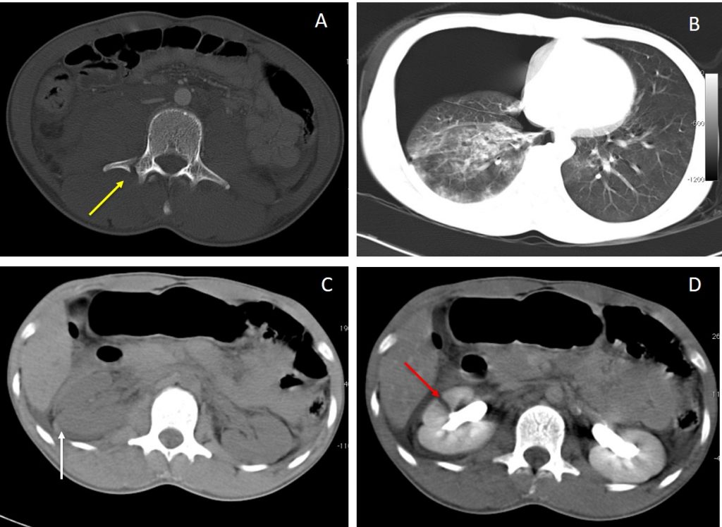
- He also sustained other injuries:

Other CT findings in this case:
- A: Bone window shows fracture of transverse process of lumbar vertebra
- B: Lung window shows right pneumothorax with lung contusion
- C: Pre contrast CT abdomen shows hyperdense collection surrounding the right renal suggestive of perinephric hematoma.
- D: Post contrast CT abdomen shows right renal laceration through the cortex not extending to the collecting system and not causing urinary extravasation in keeping with Grade III renal injury.
