Case contribution: Dr Radhiana Hassan
Clinical:
- A 49-year old single, mute and deaf
- Presented with left breast lump a few months ago, recently increased in size
- Only informed caretaker when the lesion is feels uncomfortable
- Mass felt in the left breast and enlarged left axillary nodes
Mammogram findings:
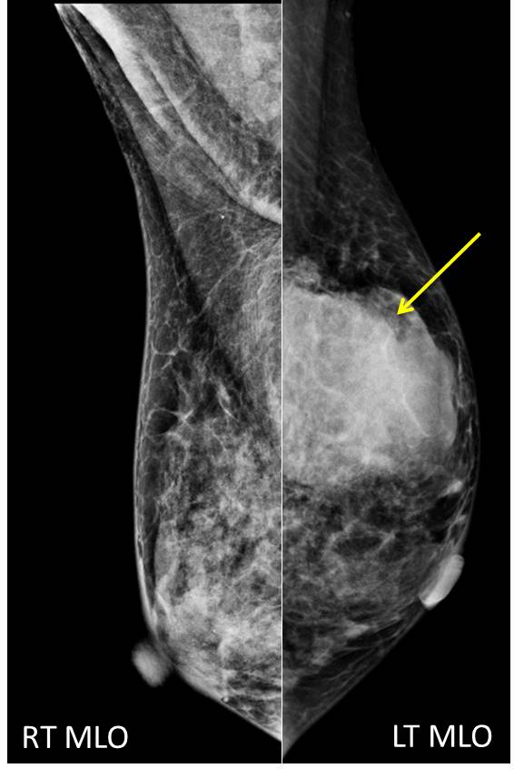
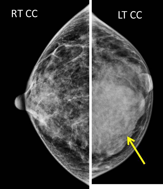
- A large mass seen at left upper inner quadrant (yellow arrows)
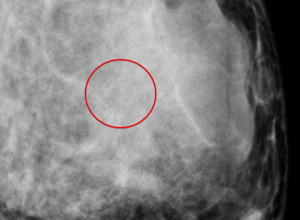
- Suspicious clustered microcalcifications are seen within the mass
- No speculated margin or architectural distortion
- No skin thickening or nipple retraction
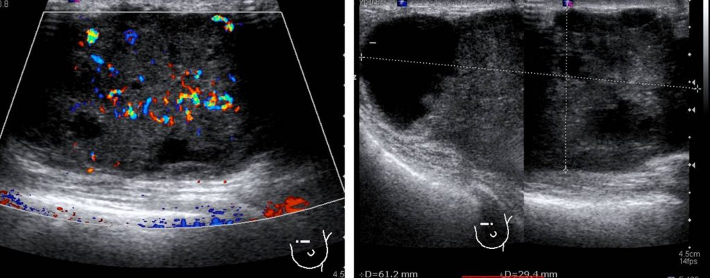
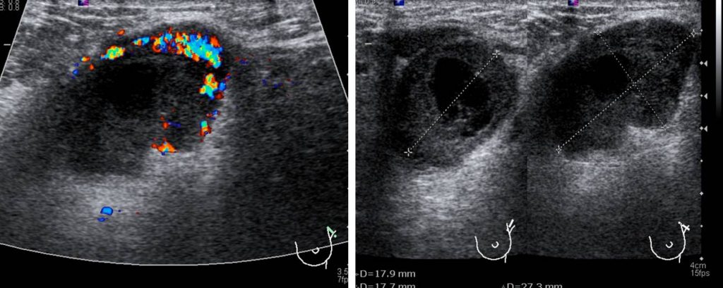
Ultrasound findings:
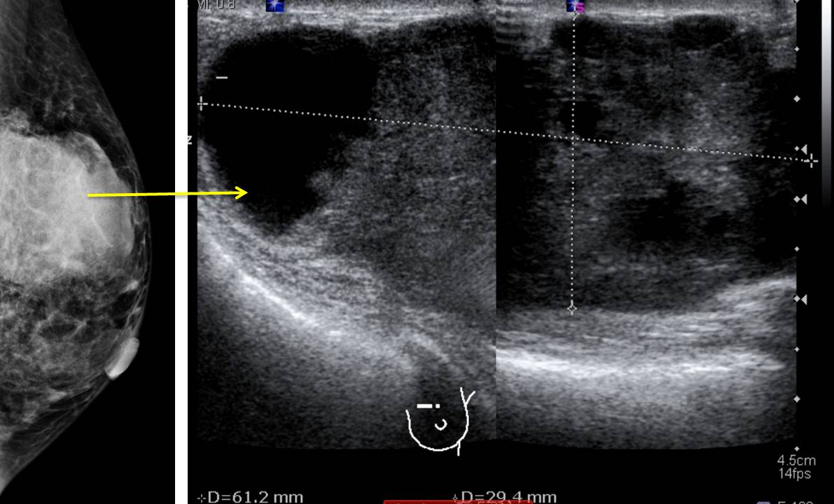
- A large mass occupying whole upper inner quadrant measuring about 6x5x4 cm.
- It shows well circumscribed border with heterogenous echogenicity
- No posterior shadowing seen
- There is increased intralesional vascularity and also presence of penetrating vessels
- A large mass at left axilla measuring about 2.0 x 1.8 x 2.7 cm.
- No other lesion in both breasts.
Progress of patient:
- Biopsy done shows invasive carcinoma
- Mastectomy done. A 70x45x35 mm mass.
- HPE: invasive carcinoma Grade III with tumour necrosis, pleomorphic nuclei and cytoplasm, no vascular invasion, one axillary node replaced by tumour cells. T3N1
- CT staging shows no distant metastasis
Diagnosis: Invasive carcinoma of the breast
Discussion:
- Invasive breast carcinoma or also known as ductal carcinoma of no special type (NOS) is the most common type of breast cancer representing 65% to 75% of cases.
- Peak age of presentation is about 50 to 60 years.
- Staging of cancer is usually done using TNM classification
- Assessment of primary tumour:
- Tx: primary tumour cannot be assessed
- T0: no evidence of primary tumour
- T1: ≤20 mm
- T2: >20mm ≤ 50 mm
- T3: >50 mm
- T4: any size with direct extension to chest wall and/or skin
