Case contribution: Dr Radhiana Hassan
Clinical:
- A 9 years old boy
- Multiple swelling at limbs
- No symptoms of compression
- No rapid progression of swelling
- Clinically presence of multiple bony-hard swelling at left elbow region, knee and proximal tibia noted.
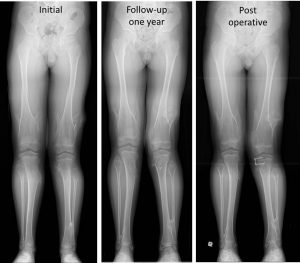
- Valgus knee left 5 degree and right 2 degree.
- Limited pronation and supination of left forearm.


Radiographic findings:
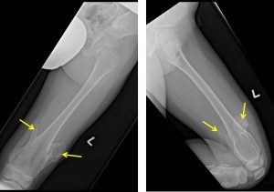
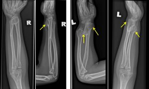
- Multiple bony projection seen distal femur, proximal tibia and distal radius ulna (worse on the left side)
- The bony outgrowth is seen projecting away from the joint line
- No cortical break is seen
- No surrounding soft tissue mass, however bulge of soft tissue seen at the region of bone outgrowth. No calcification within the soft tissue.
- No fracture.
Diagnosis: Hereditary multiple exostoses
Discussion:
- Exostoses are defined as benign growths of bone extending outwards from the surface of a bone.
- Where exostoses are capped with cartilage, they are termed osteochondromas, which can be solitary or multiple, sessile or pedunculated
- Hereditary multiple exostoses, also known as diaphyseal aclasis or osteochondromatosis.
- It is an autosomal dominant condition characterized by the development of multiple osteochondromas.
- Most patients are diagnosed by the age of 5 years, and virtually all are diagnosed by the age of 12 years.
- The skeletal distribution of lesions varies with some show typical bilateral and symmetric distribution, whereas others show a strong unilateral predominance.
- Complications include vascular impingement, neural impingement, fracture, bursitis, deformity and ankylosis.
- Malignant transformation is more common than in sporadic cases, with transformation rates reported as high as 25%.
Progress of patient:
- Worsening left genu valgus secondary to multiple exostosis
- Left medial hemiepiphysiodesis done

Reference:
- Hereditary multiple exostoses. Radiopedia at https://radiopaedia.org/articles/hereditary-multiple-exostoses
