Case contribution: Dr Radhiana Hassan
Clinical:
- A 72 years old man
- Underlying DM on insulin injection, HPT, dyslipidaemia and gouty arthritis
- Presented with pus discharge at right foot for 3 days
- It is associated with pain and fever
- Clinically right foot is dry with podagra measuring about 5×4 cm. There is surrounding cellulitis and 2 ulcers with pus discharge
- DPA and PTA arteries are palpable. Full range of motion of the ankle


Radiographic findings:
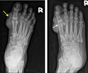
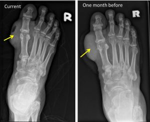
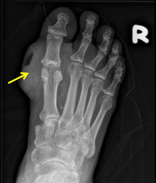
- A large soft tissue swelling adjacent to the first metatarsophalangeal joint
- It is eccenterically located with air pocket within (yellow arrow)
- Subarticular bone erosion seen of the head of adjacent metatarsal (white arrow)
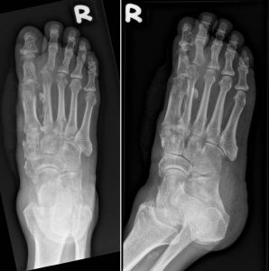
- No fracture or dislocation
Intra-operative finding
- Wound debridement done under GA
- Wound over the gouty tophi debridement and extended dorsally and plantarly until healty soft tissue seen
- Pus discharge around the gouty tophi and extending into the 1st metatarsal bone
- Head of metatarsal bone is fairly good condition with bleeder seen after removal of chalky necrotic tissue overlying it

Diagnosis: Infected tophus
Discussion:
- Tophus/tophi usually appear as lumps on the skin over affected joints in patients with longstanding high levels of serum uric acid
- It is due to deposits of monosodium urate crystals
- Tophi are a pathognomonic feature of gout.
- On radiograph, tophi are typically seen as eccentric, juxta-articular soft tissue nodules.
- Bony erosions with sclerotic margins and overhanging edges are commonly seen in their proximity
- Tophi are not be surgically removed unless they are in a critical location or drain chronically.
- Surgery is indicated for tophaceous complications, including infection, joint deformity, compression and intractable pain, as well as for ulcers related to tophaceous erosions.
- Delayed healing is noted in 50% of patients.
