Clinical:
- A 20 years old lady
- Progressive abdominal distension
- No associated loss of weight
- No bowel related symptoms
- Menses are regular


CT scan findings:
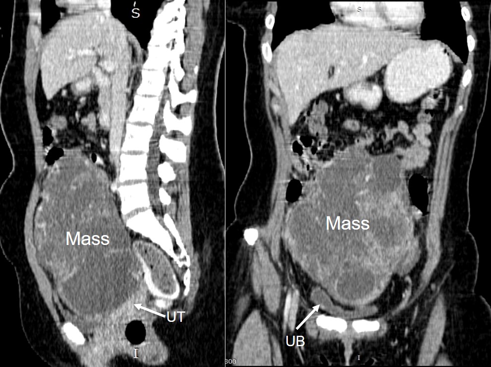
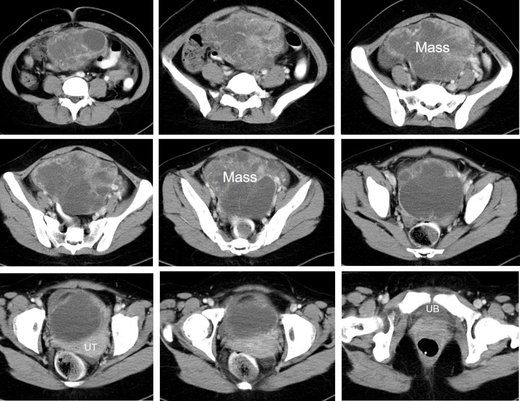
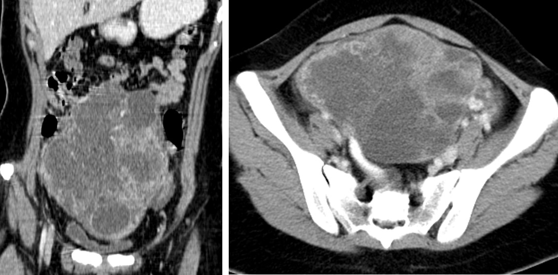
- There is a lobulated cystic mass with soft tissue component within.
- The mass measures 14x11x19 cm.
- Mass also shows thickened septa and small wall calcification.
- No fatty component is seen.
- The mass is displacing the bowel to the right side
- It causes compression to the urinary bladder and uterus
Intra-operative findings:
- Left ovarian tumour measuring about 18x20x10 cm, leaking, stuck over the right pelvic wall with suspicious invasion to the right lateral pelvic peritoneum.
- Left fallopian tube is normal.
- Uterus, right ovary and right fallopian tube are normal
- A few shorty pelvic and lower paraortic nodes.
- Minimal ascites. Other organs normal.
HPE findings:
- Macroscopy: specimen labelled as left ovarian tumour and omentum consists of a well encapsulated mass with attached fallopian tubes measuring 185x140x90 mm. The attached fallopian tube measuring 80 mm in length and 9 mm in diameter. Serial cut sections of the mass show solid cystic appearance. The solid part has grey yellow cut surface with areas of necrosis. The cystic area is variable in size ranging from 1-100 mm in diameter. The wall thickness is about 1 mm.
- Microscopy: sections from left ovarian tissue show a malignant tumour displaying vesicular, microcystic pattern composed of a loose stroma and thin walled spaces of varying sizes. The microcysts are lined by flattened to pleomorphic cells exhibiting vesicular nuclei, prominent nucleolus and clear cytoplasm. Mitosis are frequently seen. Many hyaline globules are present (PAS- positive, diastase-resistant). Schiller-Duval bodies are also noted. There are large area of necrosis and haemorrhage seen. Section from the omentum show a congested fibroadipose tissue with no evidence of malignancy. Fallopian tube is unremarkable.
- Interpretation: Left ovarian mass: Malignant yolk sac tumour.
- No malignancy in 22 lymph nodes and peritoneal wall
Diagnosis: Malignant yolk sac tumour of left ovary
Discussion:
- A yolk sac tumor is a rare, malignant tumor of cells that line the yolk sac of the embryo. These cells normally become ovaries or testes.
- A yolk sac tumour is also known as endodermal sinus tumour.
- It is a type of ovarian germ cell tumour.
- It is most often found in young children, but can occur throughout life.
- Typically, yolk sac tumours manifest as a large, complex pelvic mass that extends into the abdomen and contains both solid and cystic components.
- The cystic areas are composed of epithelial lined cysts produced by the tumour or of co-existing mature teratomas.
- Bilaterality is rare.
