Case contribution: Dr Radhiana Hassan
Clinical:
- A 2 years old boy
- Right eye leukocoria since 6 months old
- Examination shows absent reflex in right eye
- Anterior segment leukocoria with vascularization
- B scan shows lobulated lesion in right eye

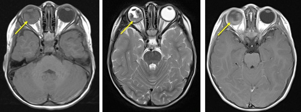
- MRI brain in axial plane, T1, T2 and T1+Gadolinium sequences
MRI findings:
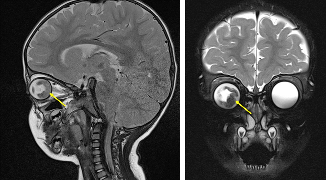
- Lobulated right retinal mass (yellow arrows)
- Measures 3.6 cm in widest length, 0.9 cm in thickness
- Intermediate signal on T1, low signal on T2
- Restricted diffusion on dwi and blooming artifact also seen (images not shown)
- Mild contrast enhancement post contrast
Diagnosis: Retinoblastoma (HPE proven)
Discussion:
- Retinoblastoma is the most common pediatric intra-ocular tumour.
- The average age at diagnosis is 18 months.
- About 80% of cases occurring before the age of 3–4 years old.
- It is a highly malignant tumour of the primitive neural retina.
- About 30% are bilateral.
- Lesions may be synchronous, metachronous, unifocal or multifocal.
- It may be endophytic, exophytic or diffusely infiltrating.
- Ultrasound shows a solid echogenic mass with high echogenic foci of calcifications.
- At T1-weighted MR imaging, retinoblastoma shows intermediate signal intensity, slightly hyperintense to the vitreous. The vitreous may be abnormally bright on T1-weighted images because of increased globulin content and a decreased ratio of albumin to globulin that occurs with malignancy.
- At T2-weighted MR imaging, the tumour is usually dark compared with the vitreous. The partially calcified areas may appear as hypointense foci within the tumour
- The tumour shows moderate to marked enhancement.
- The tumour shows restricted diffusion on diffusion weighted imaging
References/Further reading:
- Guidelines for imaging retinoblastoma: Imaging principles and MRI standardization. Paediatric Radiology 2012; available at https://www.ncbi.nlm.nih.gov/pmc/articles/PMC3256324
- Retinoblastoma at https://radiopaedia.org/articles/retinoblastoma
