Case contribution: Dr. Radhiana Hassan
Clinical:
- A 33 years old male, no known medical illness
-
Patient initially presented with status epilepticus at the age of 29 years old (2017).
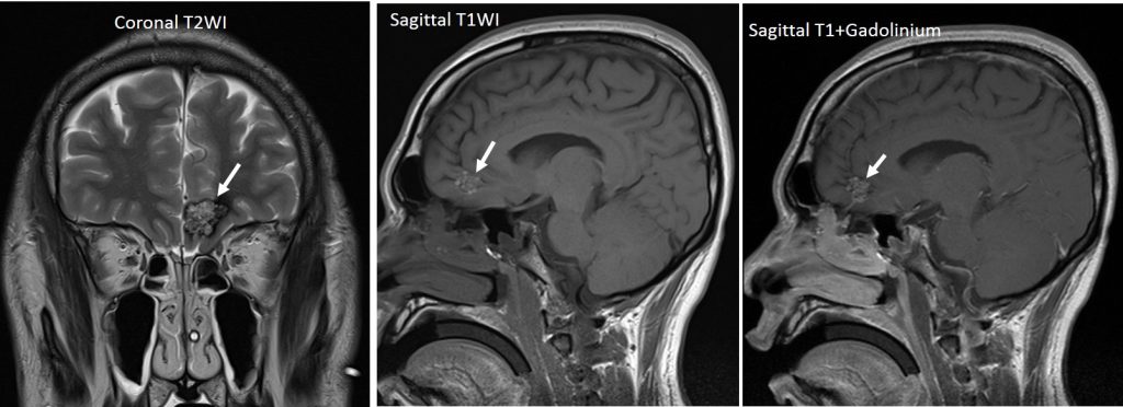
- MRI brain was done and it showed left inferior frontal lobe lesion measuring 1.7 x 1.4 x 1.4cm.
- EEG showed bifrontal spike with right side > left side.
- Patient was started on antiepileptic, however, patient was not compliance to medication and presented again with status epilepticus in year 2018 and 2019 with both requiring intubation.
- Patient has no blurry vision. No symptoms and signs of raised ICP, No loss of smell, no symptoms of frontal lobe syndrome
- Clinically higher motor function intact, MMSE 29/30, Pupils 3/3 reactive, ADL independent, Cranial nerve 1- 12 intact
- CNS examinations: Bilateral upper and lower limb power 5/5, normal tone and reflexes, No cerebellar sign


MRI findings:
- A lesion is seen in the left frontal lobe measuring about 1.7 x 1.4 x 1.4cm.
- It is heterogenously hyperintense on T2, iso with hyperintense foci on T1WI and do not enhanced post contrast. A hypointense rim seen. No significant perilesional oedema.
- No restricted diffusion. Blooming artefact seen
Diagnosis: Cerebral cavernous venous malformation
Discussion:
- Cerebral cavernous venous malformation also known as cavernomas
- It is also known as slow flow venous malformations
- It is a common cerebral vascular malformations, after developmental venous anomaly and capillary telangiectasia
- Most patients present symptomatically at 40-60 years old
- Most patient have single lesions.
- Multiple lesions may be familial
- It tends to be supratentorial but can also be found in the brainstem
- It may be difficult to see on CT scan. It do not enhance. If large area of hyperdensity due to blood product and speckles of calcification.
- MRI, T1 variable depends on age of blood, small fluid levels may be evident. T2 shows hypointense rim with variation in signal depending on age of blood product. Prominent blooming on GRE or SWI sequences.
