
Clinical:
- A 55 years old man
- Underlying hypertension and DM
- Presented with acute onset of altered consciousness
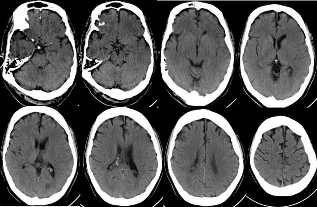
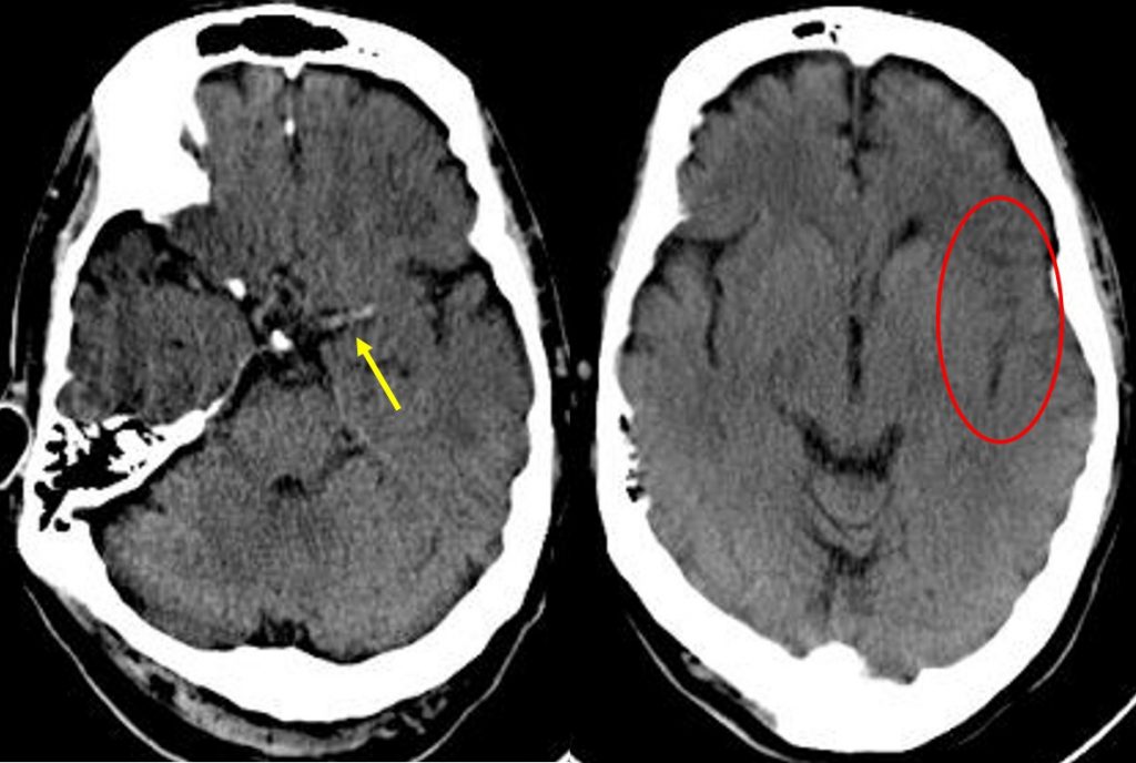
CT findings:
- There is no intracranial haemorrhage
- Hyperdense MCA sign seen on the left side (yellow arrow)
- Insular ribbon sign with effacement of left sylvian fissure
- No definite area of hypodensity is seen
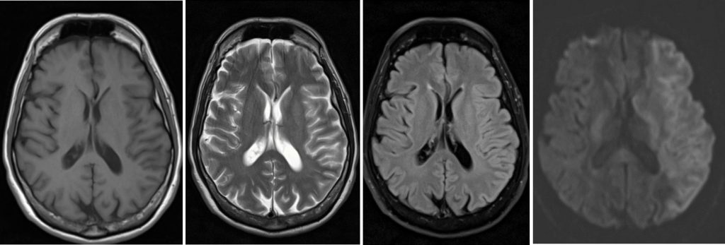
MRI findings:
- Abnormal signal intensity at left MCA territory
- Hypointense on T1, hyperintense on T2 and not well seen on FLAIR
- The region showed restricted diffusion on DWI/ADC images
Diagnosis: Hyperacute left MCA infarction
Discussion:
- The middle cerebral artery territory is the commonest affected territory in cerebral infarction
- The earliest finding in MCA infarction is hyperdense MCA sign that represent direct visualisation of thromboembolism
- Early parenchymal sign include decreased attenuation involving the lentiform nucleus, caudate nucleus and at insular ribbon region
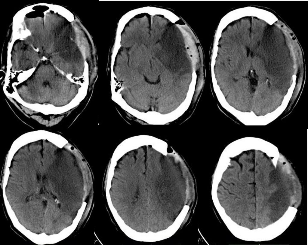
Progress of patient:
- Decompression craniectomy done
- Patient was discharged, able to ambulating but had expressive dysphasia and unable to work
Recent Comments