Clinical:
- A 35 years old man
- Involved in motor vehicle accident
- complains of painful swelling of left knee
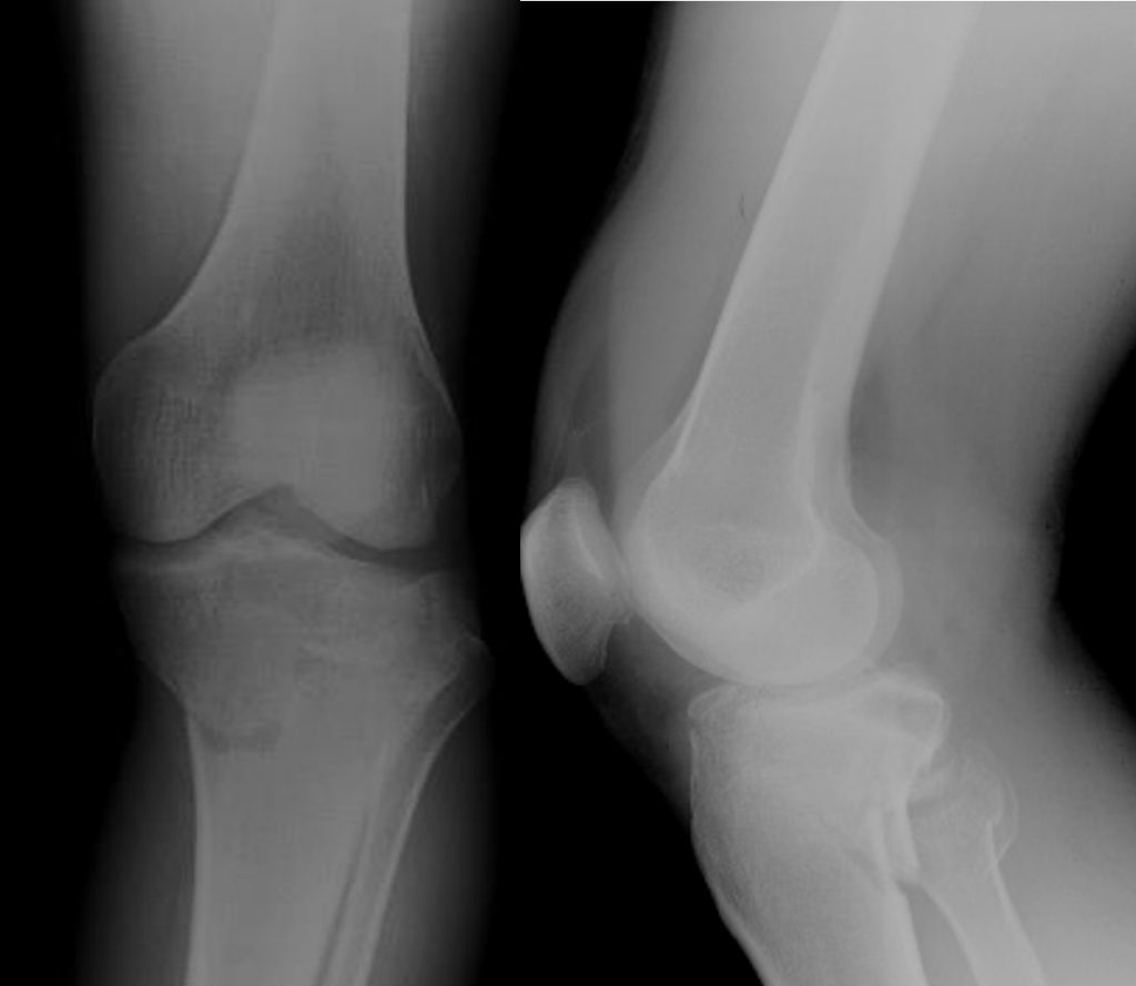
Radiographic findings:
- There is comminuted fracture involving the tibial plateau (white arrows)
- Extension of fracture lines to articular surface
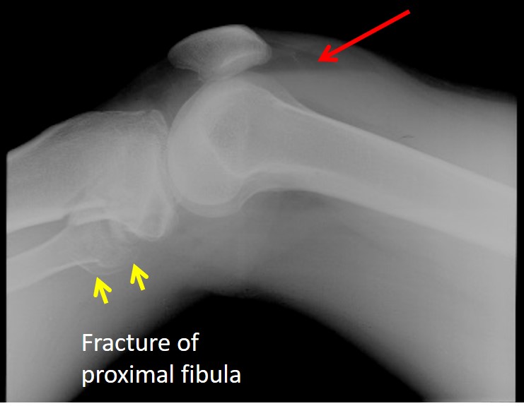
- Fracture also seen involving the proximal fibula (yellow arrows)
- There is fluid level seen at suprapatellar pouch (red arrow)
![]()
Diagnosis: Lipohemarthrosis from fracture of tibial plateau
Discussion:
- Lipohemarthrosis results from an intra-articular fracture when fat and blood from the bone marrow is extruded into the joint space.
- It is most commonly seen in the knee but has also been described in the shoulder, elbow and hip.
- On x-ray the fat-fluid level is seen with a horizontal beam. Hemorrhagic fluid is usually homogeneous. If free fat is present, the fluid is not of similar density, and will tend to separate into layers.
- This is also known as FBI sign (fat blood interface).
- The fat-fluid level may also be detected with ultrasound, CT or MRI.
- A lipohemarthrosis is significant because it is an indication of severe injury with tearing of the synovial membrane and presence of an intra-articular fracture – if a fracture is not seen on x-ray, further imaging is required.
Recent Comments