
Clinical:
- A 57-year-old man
- Presented with headache and diplopia.
- Clinical examination showed ptosis of left eye with CNIII palsy.
- There was no body weakness.
Imaging finding:
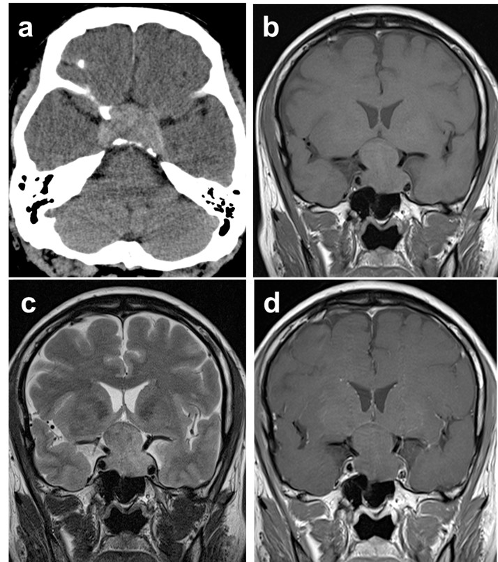
- The non-contrast axial plane soft tissue window CT scan of the brain (a) showed a hyperdense lesion at the sella region. There is no calcification within the lesion.
- Coronal MRI images (b) T1-weighted, (c) T2-weighted and (d) Post contrast image showed a sella lesion with suprasellar extension which is isointense on T1, hyperintense on T2 with minimal contrast enhancement.
- The classic ‘figure of eight’ or ‘snowman’ appearance demonstrated in this case.
- Compression and elevation of optic chiasm is also seen.
- Both carotid arteries were encased within the lesion but patent.
Diagnosis: Pituitary macroadenoma (HPE proven)
Discussion:
- Pituitary macroadenomas are the most common suprasellar mass in adults.
- They are defined as pituitary adenomas greater than 10mm in size.
- Patients typically present with symptoms of local mass effect on adjacent structures especially the optic chiasm.
- Imaging shows sellar mass without separate identifiable pituitary gland.
- CT scan shows variable attenuation sella mass usually isodense to gray matter.
- The best imaging technique for assessment is MRI with sagittal or coronal T1 and T2-weighted images. On MRI this lesion usually is isointense to gray matter on T1WI and T2WI, most lesion show moderate heterogenous enhancement post contrast but some adenomas can be hypoenhancing.
- The morphological ‘snowman’ appearance is caused by dural constriction by diaphragm sella.
- Cysts and necrosis are common occurring in about 15-20% of cases.
- Hemorrhage can occur in 10% of cases and calcification in 1-2% of cases.
Recent Comments