
Clinical:
- A 35 years old man
- Involved in MVA
- Complains of shortness of breath and chest pain
- Examination shows no breath sound at left lung
- Patient is also tachycardic, Blood pressure is normal
Radiographic findings:
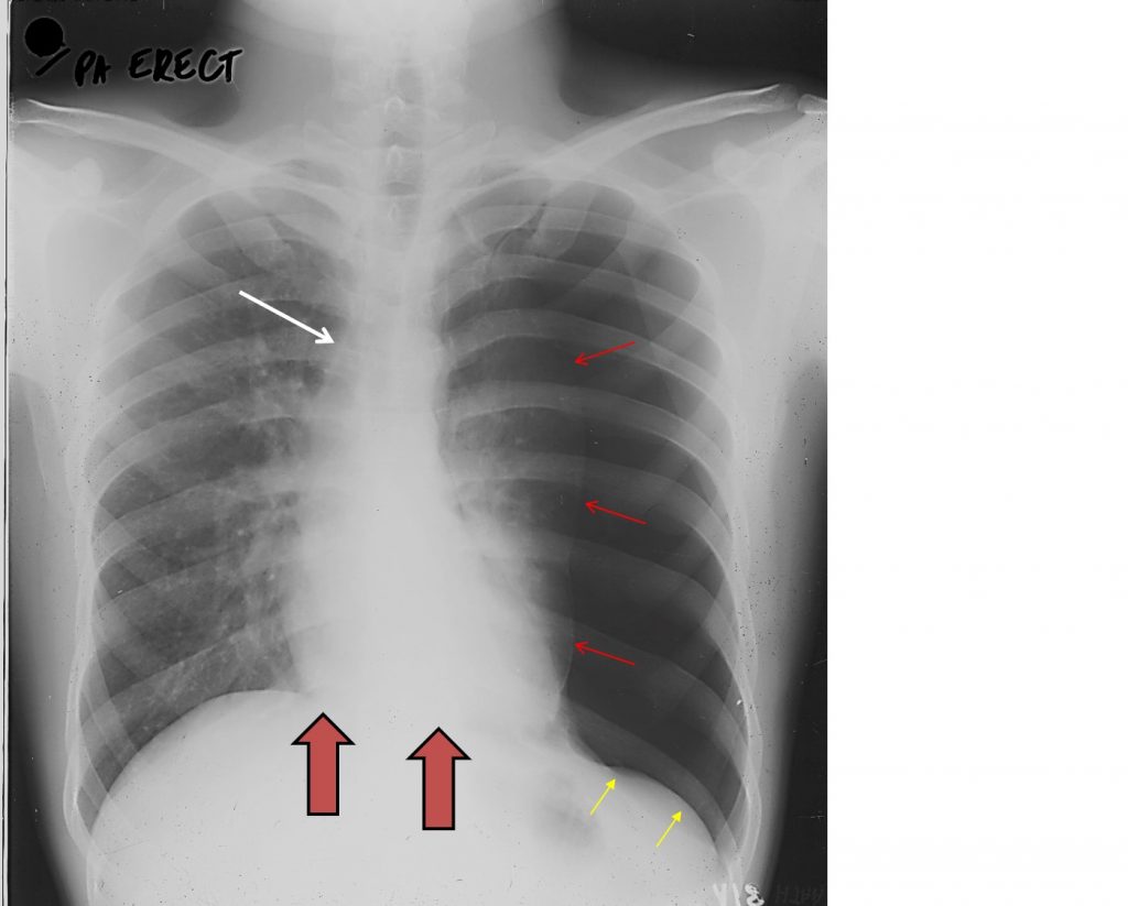
- Collapsed left lung with visualization of left pleural lining (red arrows) with no lung parenchyma distal to this
- Associated increased intercostal spaces on the same side
- Tracheal shift to the right side (white arrow)
- Depression of the left hemidiaphragm (yellow arrows)
- Shift of mediastinum to the right side (block arrows)
- No contusion in visualized aerated lung fields
- No fracture of visualized bones
Diagnosis: Tension pneumothorax
Discussion:
- Tension pneumothorax is a life-threatening condition that develops when air is trapped in the pleural cavity under positive pressure, displacing mediastinal structures and compromising cardiopulmonary function.
- Radiographic findings include the typical findings of pneumothorax with contralateral shift of mediastinum and trachea, ipsilateral depressed hemidiaphragm and increased intercostal spaces.
- Treatment of a tension pneumothorax is one of the classic medical emergencies where life can be saved or lost on the basis of recognition and subsequent rapid decompression.
- Numerous techniques exist, but in the first instance relieving the tension, even if not draining the pneumothorax, is life-saving.
- A needle thoracostomy (e.g. 14G intravenous cannula) can be inserted, typically in the 2nd intercostal space in the midclavicular line, to gain valuable time, before a larger underwater drain chest tube can be inserted.
Recent Comments