
Clinical:
- A 41 years old lady
- Post laparotomy day 2
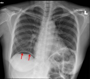
Radiographic findings:
- Free gas is seen under the dome of right hemidiaphragm (red arrows)
- Left hemidiaphragm is elevated, no free air seen under it
- The aerated lung fields are normal.
- Visualized bones are unremarkable
- No pneumomediastinum or pneumothorax
Radiologic diagnosis: Post operative pneumoperitoneum.
Discussion:
- Pneumoperitoneum should be differentiated from other types of extraluminal intra-abdominal gas.
- Causes of pneumoperitoneum include following laparotomy/laparoscopy, colonic perforation, perforated peptic ulcer, introduction per vaginal e.g douching and idiopathic.
- Free gas under the diaphrag can detect minimum 10 mls of air and 10 minutes may be needed for all gas to rise in erect /semierect position
- Post operative pneumoperitoneum occurs in up to 60% of laparotomies and 25% of laparoscopic procedures
- If the volume of gas increases then another cause of pneumoperitoneum should be sought
- If there are no features of peritonitis or other concerning features, management is conservative and the gas will gradually be reabsorbed
- Two-thirds of cases resolve within 48 hours and 97% resolve within 5 days
Progress of patient:
- Patient was managed conservatively
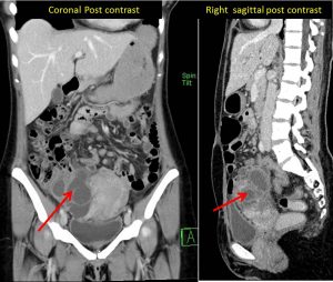
- HPE came back as adenomyosis and right endometriotic cyst
- Patient recovered uneventfully
Recent Comments