
Clinical:
- A 13 years old male
- No known medical illness
- Presented with right sided abdominal pain for 2 days and vomiting. No fever, no diarrhoea
- Hemodynamically stable
- No abdominal distension
- Clinically no mass palpable, mild tenderness at right lumbar region.
- Abdominal radiograph is unremarkable.
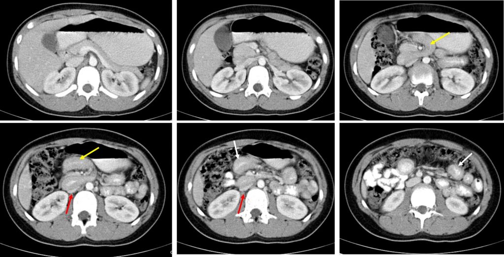
CT scan findings:
- The duodenum and proximal jejunum forms 2 loops, initially towards the right side before coursing to the left abdomen (yellow arrows)
- Thickening of bowel wall, involving the third part of duodenum extending to the proximal jejunum (white arrows)
- Mass effect with compression of IVC causing short segment narrowing of the vessel (red arrows)
- No dilated bowel loops. No extraluminal or intramural air. No ascites.
- SMA well opacified and normal in appearance. No SMA-SMV reversal seen
- Appendix, caecal pole are normal (images not shown)
Diagnosis: Midgut volvolus
Discussion:
- Potentially can occur at any age but approximately 75% of cases occur within a month of birth, 90% within a year
- Plain radiograph: non-contributory (ranges from normal to bowel obstruction to pneumoperitoneum)
- Upper GI study: corkscrew sign, tapering or beaking of bowel in compete obstruction, malrotation
- Ultrasound: clockwise whirlpool sign, abnormal superior mesenteric vessels, abnormal bowel loops, free fluid
- CT scan: whirlpool sign of twisted mesentery, malrotated configuration, inverted SMA and SMV relationship, bowel obstruction, free fluid or free gas
- Treatment: urgent surgical repair (Ladd procedure) to prevent ischaemia
Recent Comments