
Clinical:
- A 91 years old lady
- Abdominal distension for many years
- Gradual increase in size
- No constitutional symptoms
CT findings:
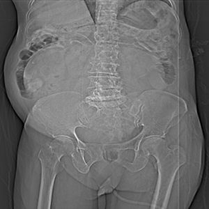
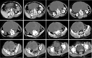
- There is a well defined cystic mass in the lower abdomen and pelvis. The mass measures 15×10 cm.
- The mass has multiple septa especially in the inferior aspect.
- There is no calcification or fat component within the mass.
- There is mass effect to the bladder and bowel. Bowel loops are displaced laterally.
- There is no ascites. No intra abdominal node enlargement.
- No hydronephrosis bilaterally.
Intra-operative findings:
- A huge left ovarian tumour of about 28 weeks size of pregnant uterus, cystic, multiseptated with thin capsule.
- No obvious solid area. Removed intact with the uterus and other adnexa.
- Right ovary is atrophic
- Both fallopian tubes are normal
- Uterus atrophic, no surface nodule seen
- Pelvis, peritoneum also no nodule. No abnormal lymph node
HPE findings:
- Macroscopy: specimen labelled as uterus, left ovary, fallopian tube and cervix. The whole specimens weighing 3600 gms. The right ovary measures 25x15x10 mm. The left ovary is grossly enlarged with smooth surface. The left ovary measures 230x240x120 mm. On cutting opened shows multilocular lesion measuring 3-100mm in diameter filled with colourless yellowish jelly like material. The cyst wall measures 2-5 mm in thickness. There is no solid area seen.
- Microscopy: section of the left ovarian cyst wall shows to be lined predominantly by a single layer of columnar epithelial cells with basally situated nuclei. In focal areas there is ‘infoldings’ of the epithelium and formation of a few glands seen. There is no cytological atypia. The right ovary and both fallopian tubes show no significant pathology. The endometrium is atrophied. The omentum is normal.
- Interpretation: Benign mucinous cystadenoma
Diagnosis: Ovarian mucinous cystadenoma.
Discussion:
- Ovarian mucinous cystadenoma principally seen in middle adult life and are extremely rare prior to menarche. The peak incidence occurs among women who are between 30 and 50 years of age.
- It is known for its potential to grow to massive proportions and are often incidentally diagnosed.
- It is typically a benign tumor accounting for 15 percent of ovarian neoplasms and up to 80 percent of all mucinous tumors.
- Ovarian mucinous cystadenoma is characteristically unilateral, only 5 percent presenting bilaterally.
- Mucinous cystadenomas are most often multilocular with thin septae. The locules may contain complex fluid, due to proteinaceous debris or hemorrhage, or both.
Recent Comments