
Clinical:
- A 72 years old man
- Chronic smoker
- Presented with chronic cough and shortness of breath
- Also complains of headache
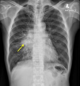
Radiographic findings:
- A suspicious ill-defined mass is seen at the right hilum measuring about 6.4 x 5.6cm.
- Non-visualization of the hilar vessels.
- No mediastinal shift. Trachea is centrally located.
- No consolidation. No pleural effusion or pneumothorax.
- Heart is not enlarged. No bony abnormality detected.
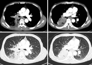
CT scan findings:
- There is a heterogeneously enhancing mass predominantly at the right superior basal segment measuring about 6.9cm (AP) x 7.4cm (W) x 6.6cm (CC).
- There is also extension into the medial segment of the right middle lobe and posterior segment of the right upper lobe.
- The mass is also seen extending to the subcarinal and left peribronchial regions. Narrowing of the branches of the right main bronchus, but no obvious endobronchial mass is observed.
- Right pleural effusion is seen, with subsegmental atelectasis of the adjacent lower lobe segments. No pleural nodule seen.
- An enlarged hilar node measuring about 1.6cm in short axis (SAD) is seen.
- Heart is not enlarged. No pericardial effusion.
Progress of patient:
- Endobronchial biopsy done shows adenocarcinoma
- CT scan brain shows multiple cerebral metastasis
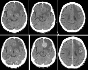
Final diagnosis: Lung adenocarcinoma with cerebral metastasis
Recent Comments