Clinical:
- A 52 years old lady
- No known medical illness
- Presented with progressive dysphagia
- Associated with significant weight loss

Fluoroscopy (Barium swallow) findings:
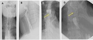
- A & B: frontal and lateral projection of upper esophagus show that the cervical part of esophagus is normal.
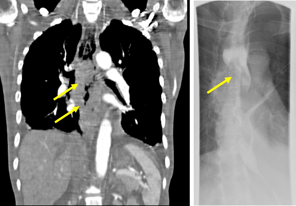
- C: Mid esophagus in frontal projection shows a lobulated intraluminal filling defect seen within the esophagus starting at the level of carina (yellow arrows).
- D: Mid and distal esophagus in frontal projection shows narrowing with minimal flow of contrast throughout the mid and distal esophagus. The GE junction appears normal.


CT scan findings:
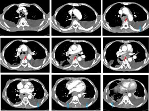
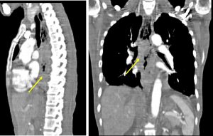
- Irregular circumferential thickening of the esophageal wall
- It involves long segment of esophagus at mid and distal portion
- Associated luminal narrowing noted at the same region (red arrows)
- The esophagus proximal to this is abnormally dilated (white arrows)
- Bilateral pleural effusion seen (blue arrows). No lung nodule.
Diagnosis: Esophageal carcinoma (HPE: moderately differentiated SCC)
Discussion:
- Esophageal cancer is the third most common gastrointestinal malignancy and is among the 10 most prevalent cancers worldwide.
- More than 90% of esophageal cancers are either squamous cell carcinomas (SCCs) or adenocarcinomas.
- SCCs are evenly distributed between the middle and lower esophagus, whereas approximately three-fourths of all adenocarcinomas are found in the distal esophagus.
- Fluoroscopy shows irregular stricture, pre-stricture dilatation with ‘hold up‘ and shouldering of the stricture.
- CT scan shows eccentric or circumferential wall thickening >5 mm, peri-oesophageal soft tissue and fat stranding. Dilated fluid- and debris-filled oesophageal lumen is seen proximal to an obstructing lesion. Tracheobronchial and aortic invasion can also be seen.

Recent Comments