
Clinical:
- A 49 years old lady
- Presented with recent onset of fitting episodes
- No neurological deficit
MRI findings:
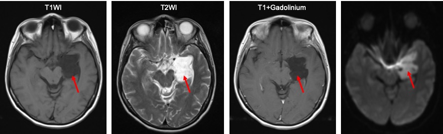
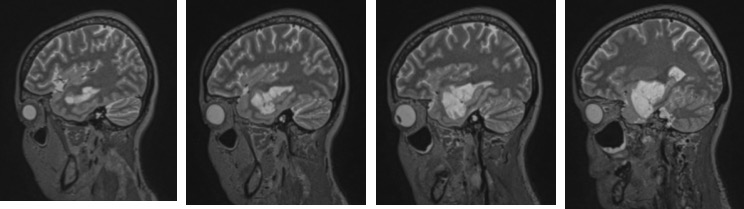
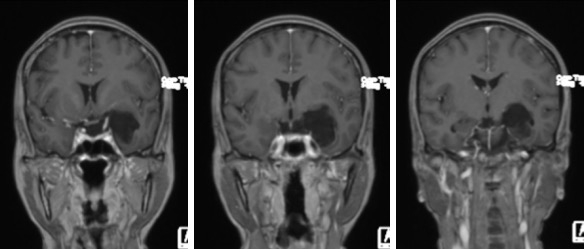
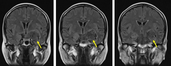
- An extra axial mass at left temporal lobe measuring 4x3x4 cm (red arrows)
- It shows intermediate signal on T1, hyperintense on T2
- Not suppressed on FLAIR (yellow arrows).
- No enhancement post contrast.
- Restricted diffusion on DWI/ADC sequences
- No extension into the cavernous sinus
- No enhancing nodule or abnormal leptomeningeal enhancement
Diagnosis: Epidermoid cyst (HPE proven)
Discussion:
- Intracranial epidermoid cyst is also known as ectodermal inclusion cyst or congenital inclusion cyst.
- It represent 0.2-1.8 % of all primary intracranial tumours
- It is the most common congenital intracranial tumours
- Symptoms depends on the site of tumour
- Location: CP angle (40-50%), 4th ventricle (17%), middle cranial fossa (10-15%)
- CT scan shows round or lobulated mass, >95% hypodense resembling CSF, 10-25% contain calcifications, no enhancement post contrast. It may cause bone erosion.
- MRI shows slightly hyperintense to CSF on T1WI, isointense (65%) to slightly hyperintense (35%) to CSF on T2WI, does not fully suppressed on FLAIR and restricted diffusion on DWI. No contrast enhancement post contrast. MRS shows no NAA, choline or lipid, resonance from lactate.
Recent Comments