
Clinical:
- A 7 years old boy
- alleged fall and left elbow pain.
- TRO left supracondylar fracture.
Radiographic findings:
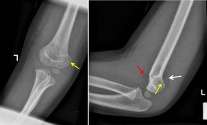
- There is supracondylar fracture with subtle fracture line seen (yellow arrows)
- The humeral condyles appear posteriorly angulated.
- Elevated anterior fat pad (red arrow).
- Presence of posterior fat pad (white arrow)
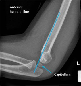
- The anterior humeral line not passing through the capitellum indicating that the condyles are displaced posteriorly.
- No dislocation of radius as evidenced by intact radiocapitellar line.
- No intraarticular loose body noted.
Radiological diagnosis: Supracondylar fracture of left humerus.
Discussion:
- Supracondylar humeral fractures are typically seen in young children, peak age of 5-7 years
- These fractures are commonly seen in boys.
- These injuries are almost always due to trauma.
- Lateral and AP radiographs are usually sufficient to demonstrate an obvious fracture.
- However, often fracture line cannot be seen in this type of fracture.
- Indirect signs of fracture include
- Anterior fat pad sig (sail sign)-anterior fat pad is elevated and appears as a lucent triangle on lateral projection
- Posterior fat pad sign
- Anterior humeral line do not intersect middle third of capitellum
Recent Comments