
Case contribution: Dr Radhiana Hassan
Clinical:
- A 45 years old man
- No known medical illness
- Presented with recurrent hematuria for few years
- No associated constitutional symptoms
- Examination shows patient had prostatomegaly
Radiographic findings:
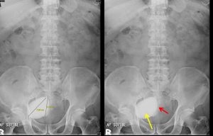
- There is an oval-shaped well-defined opacity in the pelvic region, towards the right side measuring about 7.7 cm in its largest dimension (yellow arrow).
- Another lesion seen partially superimposed with the other lesion measuring about 6.5 cm (red arrow)
- No other opacity overlying the renal region or along both ureters
- No obvious soft tissue mass lesion.
- Both kidneys and psoas outlines are clearly seen and normal.
- Degenerative changes of the spine.
- Bowel loops are grossly normal in appearance
Diagnosis: Urinary bladder calculi.
Progress of patient:
- Open vesicolithotomy done
- Intra-operative findings: Two bladder calculi seen, hypertrophic bladder wall, no obvious bladder wall growth
- Patient recovered well
Discussion:
- Urinary bladder calculi is commonly due to urinary stasis seen in bladder outlet obstruction, cystocele or neurogenic bladder
- On radiograph, it is densely radiopaque, may be single or multiple and are often large.
- On ultrasound, the calculi are mobile, echogenic with posterior shadowingy. They may be associated with bladder wall thickening due to inflammation.
Recent Comments