
Case contribution: Dr Radhiana Hassan
Clinical:
- A 46 years old lady
- Presented with right breast lump for 6 months
- Painless, progressive increase in size
- No family history of breast cancer
Mammogram findings:
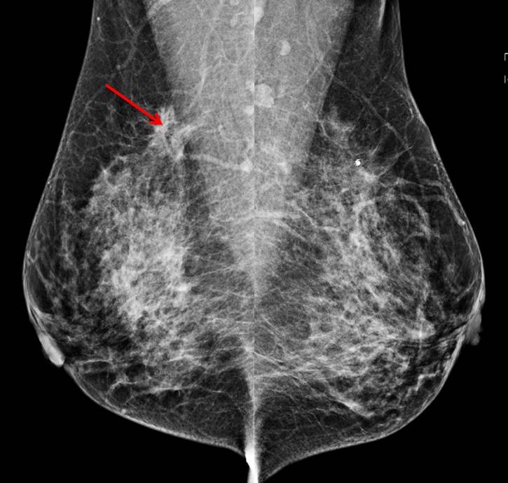
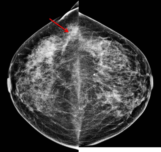
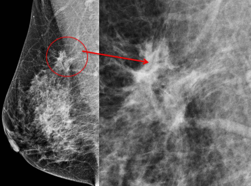
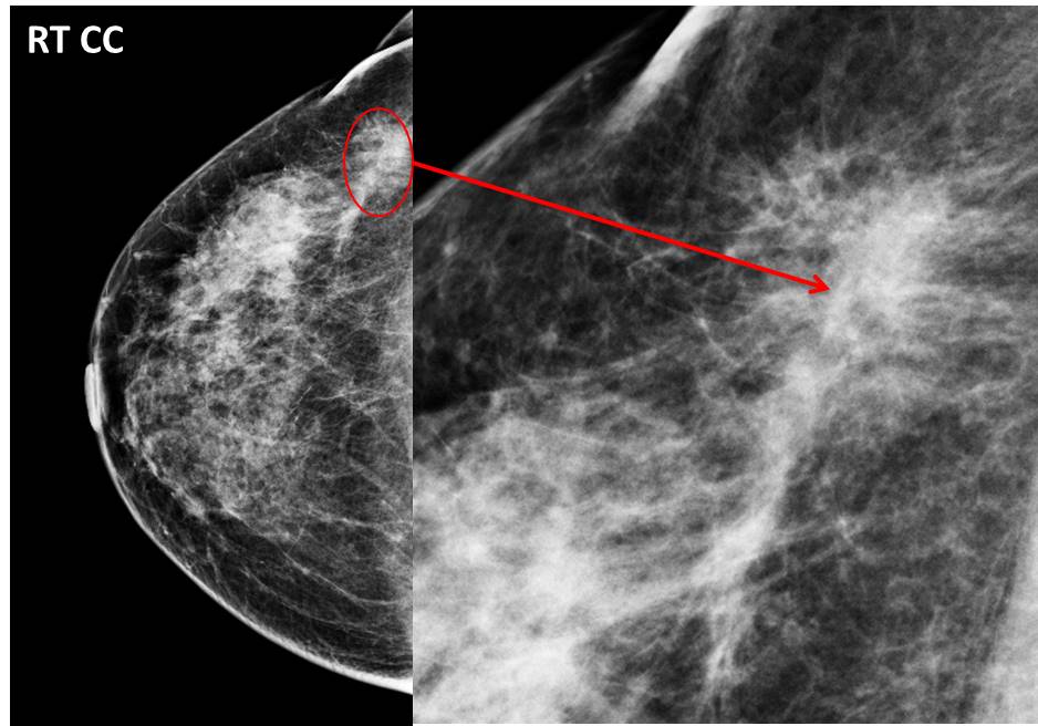
- A focal density with stromal distortion seen at right upper outer quadrant (red arrows)
- No obvious mass lesion. No suspicious clustered microcalcification
- No skin thickening or nipple retraction is seen
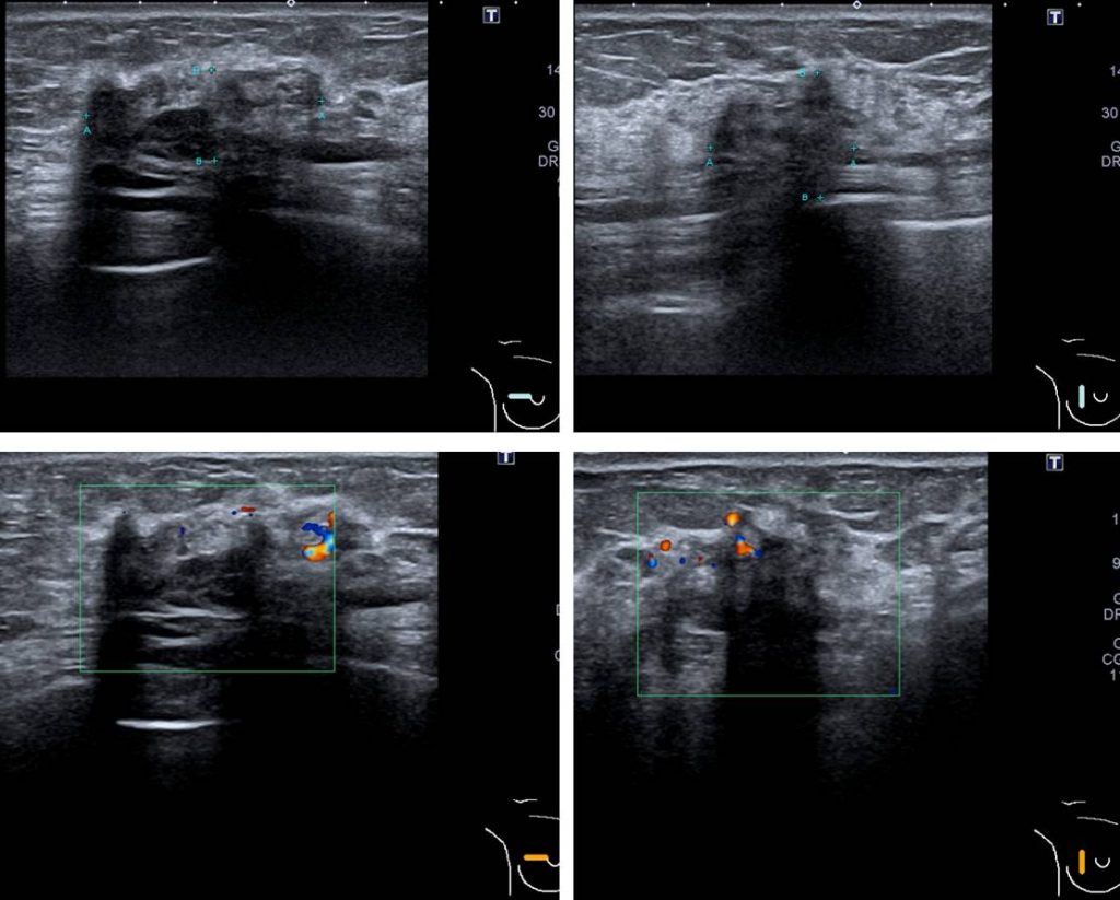
Ultrasound findings:
- An ill-defined hypoechoic mass at Rt9H-10H
- The lesion measures about 19x17x30 mm
- Posterior shadowing are seen with increased intralesional vascularity
- No ductal ectasia, no enlarged lymph nodes
Progress of patient:
- FNAC done shows presence of suspicious cells
- Biopsy done shows malignant epithelial cells in trabeculae with prominent nucleoli. ER PR +ve. Impression: invasive breast carcinoma suggestive of lobular type
- Right mastectomy and axillary clearance done one month later. HPE confirms invasive lobular carcinoma. T3N1 with 2 lymph nodes positive.
Diagnosis: invasive lobular breast carcinoma
Discussion:
- Invasive lobular carcinoma (ILC), sometimes called infiltrating lobular carcinoma, is the second most common type of breast cancer after invasive ductal carcinoma
- It accounts for about 10% -15% of all invasive breast cancers
- It begins in the milk-producing glands (lobules) of the breast.
- Compared to other types of breast cancer, lobular breast cancer:
- Has different symptoms than other more common types of breast cancer
- Often preserve the architecture of the duct which make it harder to see on mammogram. The sensitivity of mammography for the detection of Invasive lobular carcinoma reportedly ranges between 57-81%
- May not be diagnosed until the cancer is large enough to cause symptoms
- Is more likely to involve both breasts with a 5-year rate of bilateral cancer of 8% (4% synchronous and 4% metachronous tumors)
- Overall survival rate is higher than invasive ductal carcinoma
Recent Comments