
Case contribution: Dr Radhiana Hassan
Clinical:
- A 38 years old with underlying thalassemia major
- Had 3 monthly blood transfusion
- Baselin Hb=6.0 g/dL
- On iron chelation therapy
- Admitted for fever and sepsis
- Diagnosed as dengue fever (positive IgM) and meliodosis
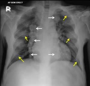
Chest radiograph findings:
- Expansion of anterior ribs at multiple levels and bilateral (yellow arrows)
- Presence of lobulated mediastinal mass with clear outline (white arrows). No calcification within.
- No lung lesion is seen.
- No cardiomegaly. No pleural effusion.
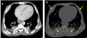
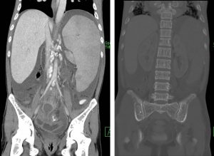
CT scan findings:
- Generalised osteopenia with irregular trabecular pattern are seen.
- Bony expansion ribs are also noted.
- Lobulated well-defined paraspinal masses are seen.
- Liver and spleen are enlarged.
Diagnosis: Extramedullary hematopoiesis
Discussion:
- Hemopoiesis is the formation and maturation of blood elements which normally occurs in the marrow of long bones, the ribs, and the vertebrae in adults.
- When the primary sites of hemopoiesis in the adult fails, various extramedullary sites take on the role of blood formation.
- Extramedullary hemopoiesis favors certain sites such as the liver, the spleen, and the paraspinal regions of the thorax.
- In the thorax, the most common imaging manifestations are paraspinal masses and rib expansion. Rib or diploic space expansion is not uncommon.
- Cases of extramedullary hemopoiesis involving the pulmonary interstitium have been reported and have occasionally resulted in cardiopulmonary insufficiency.
- Paraspinal hemopoietic tissues can extend into the central canal, especially in the thorax, and cause neurologic symptoms because of spinal cord compression.
- Diffuse hepatosplenomegaly is also a common finding.
Recent Comments