
Case contribution: Dr Radhiana Hassan
Clinical:
- A 70 years old male
- Active smoker since teenagers
- History of cholecystectomy for gallbladder stone about 20 years ago
- Presented with incomplete voiding and occasional hematuria for one month
- Also had loss of appetite and loss of weight
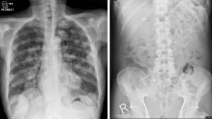
X-ray findings:
- Multiple rounded well-defined lesions are seen in both lung fields.
- These lesions are of varying sizes involving all zones of the lung.
- No cavitation or calcification within these lesions.
- No obvious bone lesion is seen in the visualized bones. No bone destruction.
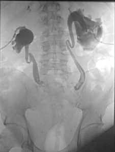
- Abdominal radiograph shows previous surgical clips, otherwise no significant finding.
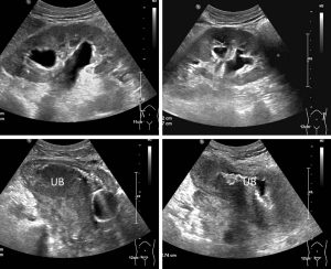
Ultrasound findings:
- Both kidneys are normal in position and echogenicity.
- There is moderate to gross bilateral hydronephrosis.
- The ureters are also dilated.
- The urinary bladder is empty with Foley’s balloon catheter in situ.
- There is diffuse thickening of urinary bladder wall.

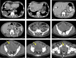
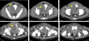
CT scan findings:
- The urinary bladder wall is grossly and irregularly thickened, which is better seen on delayed images (yellow arrows).
- Both VUJ and distal ureters are obliterated by the mass.
- Marked perivesical fat stranding is seen.
- Normal prostate and seminal vesicles are not visualised likely involved.
- Bilateral nephrostomy tubes are in situ.
- Liver is enlarged with multiple hypodense liver lesions within.
- Portal vein is patent.
- An ill-defined irregular non enhancing hypodense lesion is seen in uncinate process of the pancreas . This lesion abuts the medial wall of D2 segment of the duodenum. The rest of the pancreas is normal.
- Both adrenal glands, spleen and bowel loops are normal.
- Multiple aorto-caval and paraaortic nodes are observed.
- Multiple irregular heterogeneous masses are scattered in both lungs. The largest lesion is seen in the posterior basal segment of the right lower lobe .
Progress of patient:
- CE: huge tumour in the bladder
- TURBT: HPE shows high grade carcinoma
- Bilateral nephrostomy and internalization of both ureters done
- His condition worsened, had AKI secondary to obstructive uropathy
- Family opted for palliative treatment
Diagnosis: Cannonball lung metastases from high grade urinary bladder tumour
Discussion:
- Cannonball lung metastases refers to multiple large, well-circumscribed, round pulmonary metastases like cannonballs
- Metastases with such appearance are classically secondary to renal carcinoma and choriocarcinoma. Other primary include carcinoma of prostate, endometrium, synovium and adrenal.
- Other differential diagnosis for cannonball pulmonary lesion include various infection (septic emboli, multiple abscesses, tuberculosis, nocardia, histoplasmosis, coccidioidomycosis and hydatid cysts), rheumatological disease (Wagener granulomatosis, rheumatoid nodules) and arteriovenous malformations
Recent Comments