
Case contribution: Dr Radhiana Hassan
Clinical:
- A 40 years old lady
- Mother had breast cancer at 44 years old
- Came for screening mammogram
- Menarche at 12, no history of taking OCP
- Breast fed all 3 children about 2 years each
- Clinically no lump palpable
Mammogram findings:
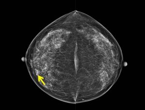
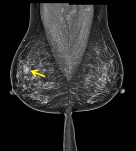
- Moderately dense breasts (Birads b) with symmetrical parenchymal pattern.
- There is no dominant mass lesion seen.
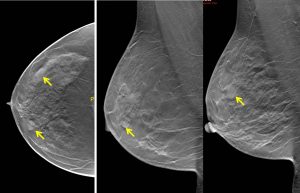
- However two focal densities are seen in the right breast (yellow arrows).
- Tomosynthesis showed well-defined lesions at this region.
- There is no suspicious clustered microcalcification.
- No stromal distortion, skin changes or nipple retraction.
- No abnormal axillary node.
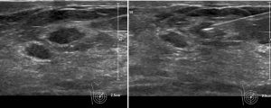
Ultrasound findings:
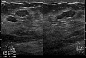
- There are hypoechoic lesions seen at Rt2H about 4 cm from nipple.
- The lesions measured 10×4 mm and 7x3mm.
- No increase in vascularity, posterior shadowing in these lesions.
- A well-defined oval-shaped lesion is seen at Rt10H measuring about 12×7 mm. No suspicious feature of this lesion.
- Small cysts seen in both breasts. No dilated duct or abnormal axillary nodes bilaterally.
FNAC done:
- Colourless fluid aspirated
- Low to moderate cellularity
- Smears composed of small mono layered sheets and clusters of bimodal populations of benign ductal epithelia and myoepithelial cells. The cells display round nuclei, regular chromatin, insconspicious nucleoli, smooth nuclear membrane, and ample cytoplasm. Occasional bare nuclei are seen in the background. Some stromal fragments are noted. No atypical or malignant cells are seen.
- Impression: Benign proliferative epithelial lesion, favours fibroadenoma
Hookwire localisation and wide local excision done:
- Macroscopy: cut section shows two small whitish tumours at the tip of hookwire measuring 10x8x8 mm and 7x6x8 mm.
- Microscopy: sections show two fairly circumscribed lesions displaying epithelial component arranged in tubules and slit-like pattern embedded within a hyalinized stroma. The glands are lined by double layer epithelium highlighted by p63 antibody. Adjacent benign breast acini are present. There is no in-situ component or invasive carcinoma seen.
- Intepretation: Features are suggestive of fibroadenomatoid nodules.
- Comment: the lesion is completely excised.
Diagnosis: Fibroadenomatoid nodules
Discussion:
- Fibroadenomatoid nodule is an uncommon lesion with histologic features similar to that of fibroadenoma but lacking well-defined borders
- Also known as fibroadenomatous change, fibroadenomatoid hyperplasia, fibroadenomatoid mastopathy, fibroadenomatosis, sclerosing lobular hyperplasia
- This lesion is distinct from the typical well circumscribed fibroadenoma that may have fibrocystic changes.
- Mean age: 28-34 years, represent 5-7% of non-neoplastic surgical biopsies,
- May present as palpable lump or incidental finding
- Lesion with features resembling a fibroadenoma but lacking sharp circumscription, often multifocal, in background of fibrocystic changes
- It is due to proliferation of intralobular stroma with the formation of stromal nodularity that oftern appears to blend in with the surrounding breast tissue, may represent a stage in evolution of fibroadenoma, produced by coalescence of fibroadenomatoid nodules
- Most common site is upper outer quadrant
- No risk of malignancy
Recent Comments