
Case contribution: Dr Radhiana Hassan
Clinical:
- A 13 years old girl
- History of pilocytic astrocytoma grade 1,
- Tumour resection done 5 years ago
- On annual surveillance MRI
- Noted to have increasing tumour size on last scan
- Otherwise patient is asymptomatic
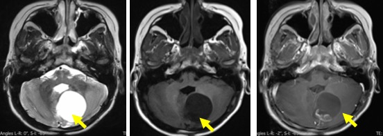
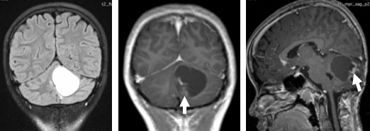
MRI findings:
- A predominantly cystic lesion is seen in the left cerebellum
- It is hypointense on T1, hyperintense on T2 and shows minimal wall enhancement
- Soft tissue component noted at its posterior wall which enhances post contrast
- No calcification or bleed within the lesion
- No surrounding oedema
- No internal brain herniation
Diagnosis: Pilocytic astrocytoma (HPE proven)
Discussion:
- Represent about 30% of paediatric gliomas
- Second most common paediatric brain tumour
- Indolent and slow growing
- Location: optic chiasm>cerebellum>brain stem
- Cerebellar lesion is usually cystic with intense mural nodule enhancement
- Calcification seen in 10% of cases
- There is strong association with NF1
Recent Comments