
Contribution: Dr Radhiana Hassan
Clinical:
- A 63 years old male
- Underlying DM, HPT and CVA
- Presented with left thigh swelling for 9 months, increasing in size
- Associated with loss of weight and loss of appetite
- No history of trauma, no history to suggest infection
- Clinical examination shows left thigh swelling 25×27 cm, non-tender, non-mobile, non-pulsatile.
- Hip and knee joint motion is full. No inguinal lymph node palpable.

Radiograph of left femur:
- A soft tissue swelling seen at left thigh region
- Associated calcification seen within it (yellow arrows)
- Otherwise no lytic or sclerotic changes of the femur
- No fracture
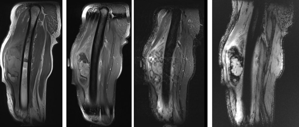
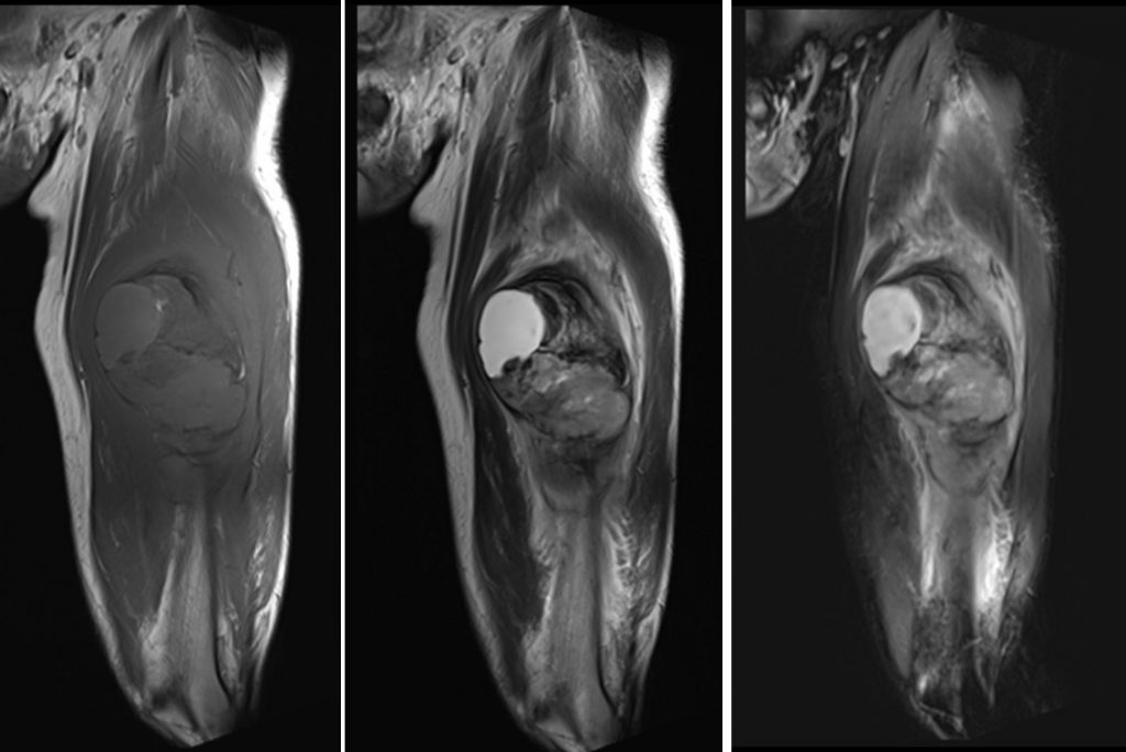
MRI findings:
- A large lobulated soft tissue mass is seen within the anterior compartment of the mid-thigh.
- The bulk of the mass is located within the vastus intermedius muscle and measures approximately 4.7 x 7.3 x 11.8 cm (AP x W x CC).
- It consists of mixed cystic and solid components and demonstrates heterogeneous hypo- to isointense on T1 and hyperintense on T2.
- A focal area of T1 hyperintensity and T2 hypointensity is seen at the superomedial aspect of the mass, most likely early subacute bleed. Late subacute bleed is seen at the inferior aspect of the mass, represented by hyperintense signal on T1 and T2.
- There are blooming artifacts seen within the mass; of which in correspondence to the previous radiograph, most likely represent calcifications rather than hemosiderin deposition.
- Restricted diffusion is noted on DWI/ADC suggesting hypercellularity.
- Post IV Gadolinium, heterogeneous enhancement of the solid component and peripheral enhancement of the cystic component is observed.
- No evidence of marrow oedema. The femoral cortex is intact. No periosteal reaction.
Progress of patient:
- Tumour excision was done
- HPE: Extraskeletal osteosarcoma
- PET scan shows FDG hypermetabolism at left medial thigh could be post-operative change or residual tumour
- Completed chemotherapy
- His condition complicated with sepsis post chemotherapy.
Diagnosis: Extraskeletal osteosarcoma (ESOS)
Discussion:
- Extraskeletal osteosarcoma (ESOS) a rare mesenchymal malignant tumour
- It is located within soft tissue without attachment to bone/periosteum.
- It is 1% of soft tissue sarcomas
- Mean age 50 years, commonest location is lower extremity in thigh (42-47%)
- History of trauma, myositis ossificans or previous site of muscular injection
- Often deep seated, average size of 9 cm
- Local or massive area of mineralization seen in > 50% of cases
- Radiograph shows ESOS as soft tissue density with variable amount of calcification that represents osteoid matrix formation.
- CT is best to demonstrate the calcification and tumour have a pseudocapsule.
- Extensive central mineralization without peripheral well-defined calcification distinguishes ESOS from myositis ossificans.
- MRI shows well-circumscribed mass with hemorrhage and necrotic areas. On T1WI it is isointense to muscle and hyperintense on T2WI.
- It shows increased uptake on bone scan
- Multiple local recurrences in 80-90%
- Metastasis to lungs, lymph nodes and bone
Recent Comments