
Clinical:
- A 35 years old man
- Known case of DM, HPT
- Also previously diagnosed as retroviral +ve
- Presented with sudden onset of headache and generalized body weakness
- Clinical examination shows GCS 15/15, no muscle weakness
CT findings:
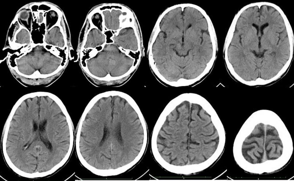
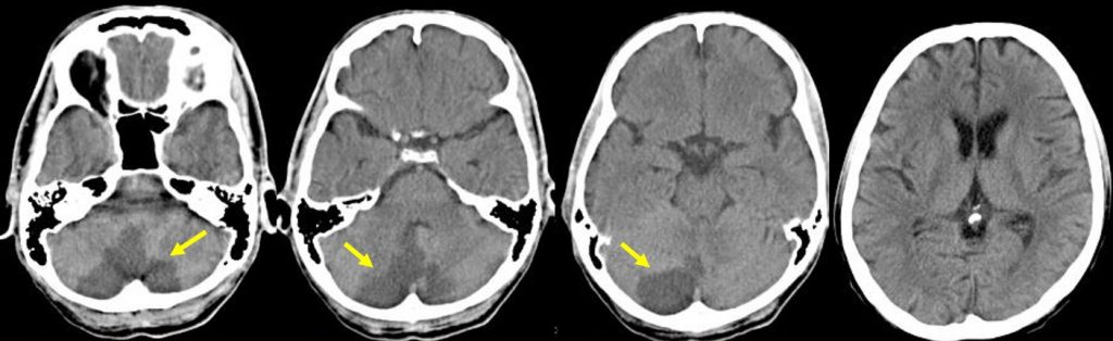
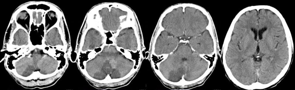
- Initial CT brain done on admission shows no significant finding
- A repeat CT brain with contrast done 2 days later
- The CT scan show hypodensity at the posterior inferior part of both cerebellum
- It is well-defined and do not show enhancement post contrast
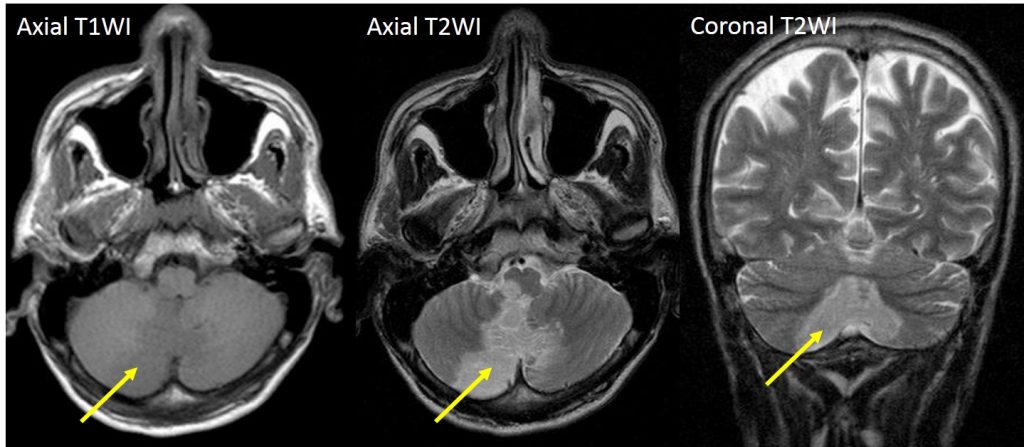
MRI findings:
- The cerebellar changes seen as hypointense on T1, hyperintense on T2 and FLAIR (yellow arrows)
- Involving posterior and inferior cerebelli, more on the right side
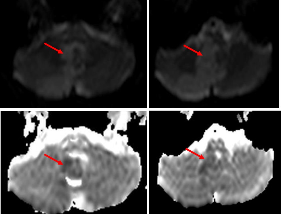
- It shows restricted diffusion
- No significant mass effect is seen
Diagnosis: PICA (posterior inferior cerebellar) infarction.
Discussion:
- Cerebellar infarction is relatively uncommon and account for about 2% of all cerebral infarction
- PICA is one of the three main arteries that supply the cerebellum
- PICA is the largest branch of vertebral artery and supplies posterior inferior cerebellum, inferior cerebellar vermis and lateral medulla
- Vertigo, nausea and truncal ataxia are the most common presenting features
- MRI is far superior to CT in the sensitivity of acute ischaemic stroke across all vascular territories.
- In the acute phase T2WI will be normal, but in time the infarcted area will become hyperintense.
- The hyperintensity on T2WI reaches its maximum between 7 and 30 days. After this it starts to fade.
- DWI is already positive in the acute phase and then becomes more bright with a maximum at 7 days.
- DWI in brain infarction will be positive for approximately for 3 weeks after onset
- ADC will be of low signal intensity with a maximum at 24 hours and then will increase in signal intensity and finally becomes bright in the chronic stage.