
Clinical:
- A 39 years old lady
- Presented with left eye symptoms
- Imaging showed lesion at left cavernous sinus area
- Biopsy done showed inflammatory cells
- On regular follow up since then
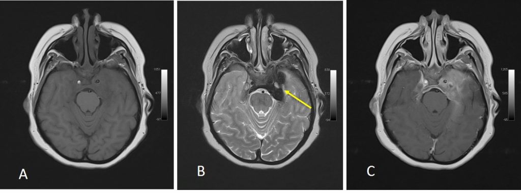
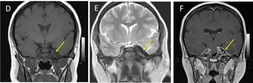

MRI findings:
- There is lesion seen at the floor of middle cranial fossa involving the sella and cavernous sinus, more on the left side (yellow arrows).
- It is hypointense on T1 and T2WI with avid contrast enhancement.
- Diffuse smooth pachymeningeal thickening and enhancement extends along the left internal auditory canal and along the prepontine cistern ending at the mid clival region (white arrows).
Diagnosis: Inflammatory pseudotumour of skull base
Discussion:
- It is a benign, idiopathic disease that is often mistaken for a neoplasm or infection owing to its aggressive behavior and clinical presentation.
- Infiltrating lesion can be seen at intraorbital, cavernous sinus, meningeal, skull base and nasopharynx
- It is seen as enhancing soft tissue mass, with lesion typically iso to hypointense on T2WI
- Bone changes are unusual
- When involving cavernous sinus, ica narrowing always present
- As in this case, tissue biopsies reveal acute or chronic inflammation without evidence of malignant disease or infection.
Recent Comments