
Clinical:
- A 45 years old man
- Alleged involved in motor vehicle accident
- Complains of shortness of breath
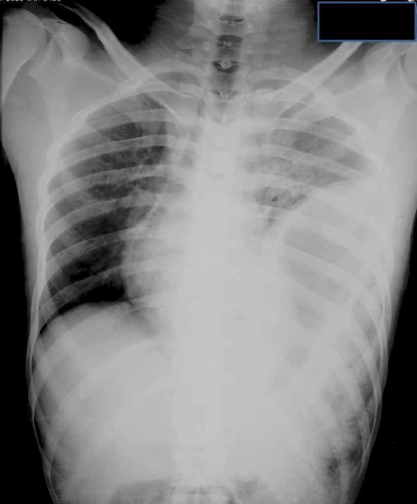
Radiographic findings:
- There is herniation of the stomach into left thoracic cavity (red arrow)
- The left hemidiaphragm is not seen
- There is shift to mediastinum and trachea (white arrow) to the right side
- Presence of collar sign (yellow arrow)
- Left lung shows compression effect
- No pneumpthorax. No rib fracture.
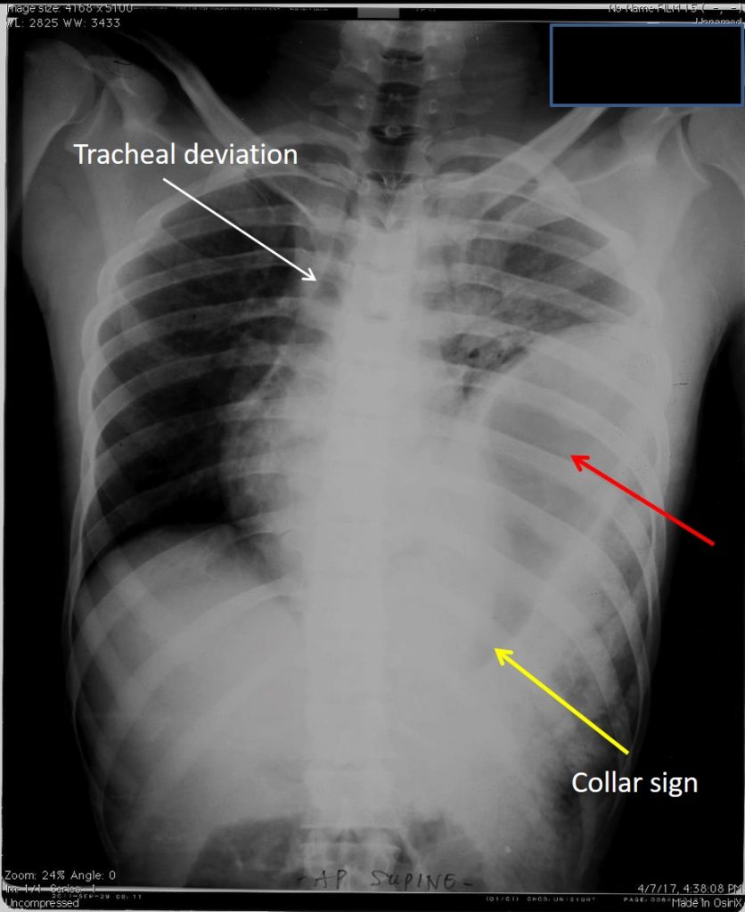
Diagnosis: Traumatic diaphragmatic hernia
Discussion:
- Traumatic diaphragmatic hernia often occur after a motor-vehicle collision. The estimated incidence ranges from 0.8-8% of patients who sustain blunt abdominal or lower thoracic trauma
- Blunt diaphragmatic injuries with intrathoracic herniation of abdominal viscera carry a high mortality rate from 30 to 80%.
- The herniation of the viscera through the rent in the diaphragm results in respiratory compromise. Visceral incarceration may occur with or without strangulation or perforation.
- The left hemidiaphragm is involved three times more frequently than the right.
- The most common site of rupture is the posterolateral aspect of the hemidiaphragm between the lumbar and intercostal muscle slips.
- Ruptures occur radially and most are >10 cm in length 4. The most commonly herniated viscera are the stomach and colon.
- inability to trace the normal hemidiaphragm contour
- intrathoracic herniation of a hollow viscus
- focal constriction of the viscus at the site of the tear (collar sign)
- if large, the positive mass effect may cause a contralateral mediastinal shift
- visualization of a nasogastric tube above the hemidiaphragm on the left side
- left hemidiaphragm much higher than the rightSpecific diagnostic findings of diaphragmatic rupture on chest radiographs may not be seen in up to 50% of cases. However, the following signs are helpful in making the diagnosis
- Many different modalities including conventional radiography, fluoroscopy, CT, and magnetic resonance imaging (MRI) have been used to evaluate the diaphragm;
- Currently, multidetector CT (MDCT) is the modality of choice for detection of diaphragmatic injury with a sensitivity and specificity of 61-87% and 72-100%, respectively.
Recent Comments