
Clinical:
- An 11 years old boy
- Alleged fall from double decker bed
- No loss of consciousness
- Had a few episodes of vomiting after fall
- No limb weakness, no headache, no blurred vision
- GCS on arrival to ED is 15/15, no neurological deficit
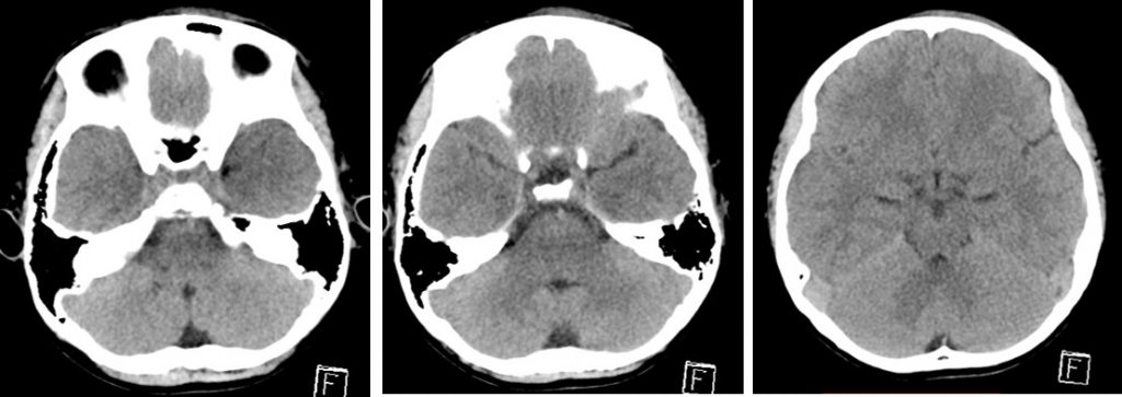
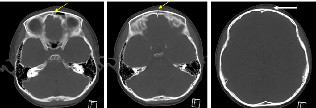
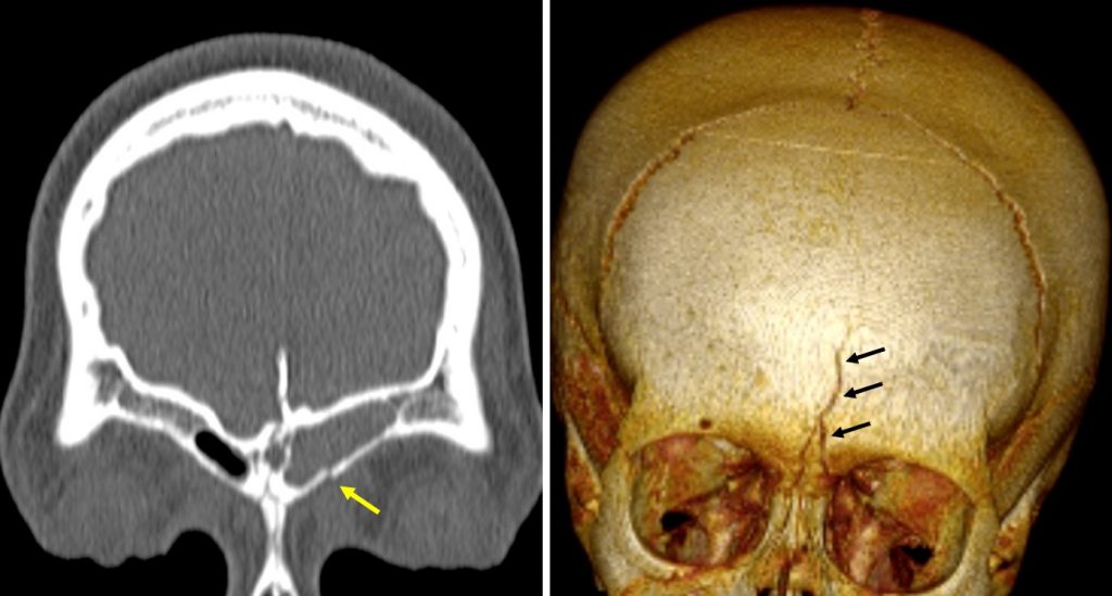
CT findings:
- There is fracture involving the left frontal bone (yellow arrows)
- It involves anterior wall of left frontal sinus, no extension to posterior wall of the sinus
- No displacement of fracture fragment
- Blood collection is seen in the left frontal sinus
- Scalp hematoma at the fracture site (white arrow)
- No intracranial hemorrhage, no pneumocephalus
Diagnosis: Linear skull fracture of left frontal bone
Discussion:
- Plain radiographs have a limited role in diagnosing skull fracture.
- Skull fractures are best imaged with CT of the brain. CT is sensitive to detect fractures also able to exquisitely characterize their extent and allow for surgical planning. Furthermore, it allows assessment of the brain parenchyma at the same time.
- Fractures will appear as discontinuities in the bone and may or may not be displaced.
- They need to be distinguished from normal sutures, which have corticated margins that fractures lack.
- If the fracture involves a paranasal sinus, middle ear or mastoid air cells, then they will contain some blood, which is a helpful clue to the presence of an underlying fracture.
- Skull fracture is absent in ¼ of fatal injuries at autopsy.
- Skull fractures are associated with 15% concomitant C-spine injury.
Progress of patient:
- Patient was managed conservatively.
Recent Comments