
Clinical:
- A 23-year old lady
- No known medical illness
- Presented with chronic headache.
MRI findings:
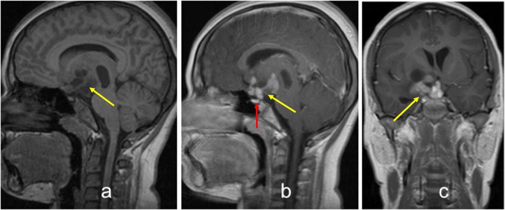
- A lobulated suprasellar mass (yellow arrows).
- It has mixed cystic and solid components.
- The lesion show marked enhancement after contrast administration.
- There is no invasion into the pituitary fossa.
- The pituitary gland is identified, normal in appearance and separated from the lesion (red arrow).
Diagnosis: Suprasellar germinoma (HPE proven)
Discussion:
- Intracranial germinomas are a type of germ cell tumour
- It is also known as dysgerminomas or extragonadal seminomas.
- These lesions are predominantly seen in paediatric patients. Peak age of incidence is 10-12 years of age with 90% of patient being younger than 20 at the time of diagnosis.
- They tend to occur in the midline either at the pineal region or along the floor of the third ventricle/suprasellar region.
- Suprasellar germinoma is more frequently seen in female.
- On CT, the lesion is slightly hyperdense compared to the adjacent brain with marked enhancement post contrast. Cystic components are found in up to 45% of cases.
- MRI demonstrates a lobulated soft tissue mass; isointense to slightly hyperintense on T1 and T2-weighted images. It may have areas of cyst formation, hemorrhage, central calcification and have predilection to cause adjacent brain oedema. It shows vivid and homogenous enhancement post contrast.
Recent Comments