
Clinical:
- A 39-year old male
- History of epilepsy since 9-year old.
- Recently fits becoming more frequent and not responding to medication.
MRI findings:
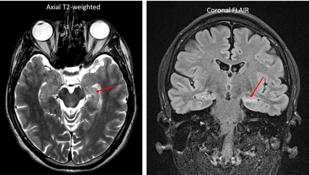
- The left hippocampus is significantly smaller that the right side (red arrows)
- It also shows hyperintense signal on T2/FLAIR images.
- No other abnormality in the rest of brain parenchyma.
Diagnosis: Mesial temporal sclerosis.
Discussion:
- Mesial temporal sclerosis is also known as hippocampal sclerosis or Ammon horn sclerosis.
- The hall mark of mesial temporal sclerosis on MRI is an atrophy of hippocampus associated with hyperintense signal on long-repetition-time sequences confined to hippocampus.
- These changes occur due to neuronal loss and gliosis. Secondary signs of this condition include ipsilateral atrophy of fornix and mamillary body, temporal lobe volume loss, narrowed collateral white matter and enlarged temporal horn of lateral ventricles.
- The findings can be bilateral in 20% of cases.
- In cases of bilateral hippocampal abnormalities, secondary findings can determine the more important side to resect.
Recent Comments