
Case contribution: Dr Raja Rizal Azman
Clinical:
- A 40-year old man
- No known medical illness before
- Presents with three episodes of tonic clonic seizures over a period of two weeks with associated loss of consciousness.
- There was no residual weakness following resolution of seizure activity.

Imaging findings:
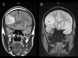
- A mass is seen in the right frontal lobe with cortical involvement appearing heterogeneously hyperintense on T2-weighted images (A) with areas of enhancement post gadolinium centrally (B).
- The lesion exhibits heterogemous hyperintense signal on FLAIR (C).
- T2*GRE sequences show focal areas of low signal within with blooming artefact (D).
Diagnosis: Oligodendroglioma (HPE proven)
Discussion:
- Oligodendrogliomas are the third most common glioma accounting for 5-18% of all glial neoplasms.
- The commonest presentation is with seizures with a peak incidence occuring in the 4th and 5th decades.
- A small proportion occurs in children.
- The frontal lobe is the commonest location (50-65%) followed by the temporal (47%) and parietal lobes (7- 20%).
- On CT oligodendrogliomas are usually heterogenously iso to hypodense. A large proportion (70-90%) will exhibit calcifications. Half of the lesions will exhibit enhancement.
- MRI typically shows a lesion appearing hypointense on T1 and hyperintense on T2. There is usually very little associated perilesional oedema. Areas of low signal on T2 and T2* sequences correspond to calcification. As on CT, 50% of these lesions will exhibit heterogenous enhancement.
- The presence or absence of enhancement has not been shown to be a reliable indicator of tumor grade.
Recent Comments