
Clinical:
- A 26 years old man
- Non-smoker with no previous medical illness
- Presented with chronic cough and constitutional symptoms
- Completed TB vaccination
Radiographic findings:
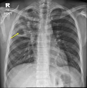
- A large cavitating lesion with surrounding consolidation is seen at the lower part of right upper lobe.
- There are multiple small cavitating lesions involving the rest of right upper lobe.
- No hilar mass. No mediastinal shift.
- Other part of the lungs are clear.
- No pleural effusion or pneumothorax. No hilar mass.
- Cardiothoracic ratio is normal.
- Bones and soft tissues are unremarkable.
Diagnosis: Pulmonary tuberculosis (sputum positive).
Discussion:
- Tuberculosis is an airborne infectious disease caused by Mycobacterium tuberculosis
- Cavitation is uncommon in primary tuberculosis, seen in only 10-30% if cases
- Post-primary infections are far more likely to cavitate than primary infections. In the vast majority of cases, they develop in the posterior segments of the upper lobes (85%).
- However, more typical appearance of post primary tuberculosis is that of patchy consolidation or poorly defined linear and nodular opacities.
- Hilar nodal enlargement is seen in only approximately a third of cases.
- Lobar consolidation, tuberculoma formation and miliary TB are also recognised patterns of post-primary TB but are less common.
Progress of patient:
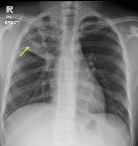
- Patient shows good response to treatment clinically
- A repeat chest radiograph after two months of medical treatment shows reduction in size of the cavity
Recent Comments