
Clinical:
- A 66-year old man
- Underlying hypertension, otherwise no other medical problem
- Presented with left eye proptosis with infraorbital swelling for 10 years.
- Gradually increase in size
- Scope shows mass lesion at roof of ethmoid sinus
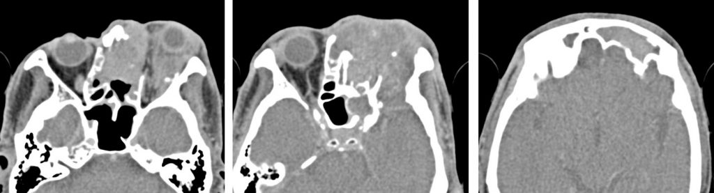
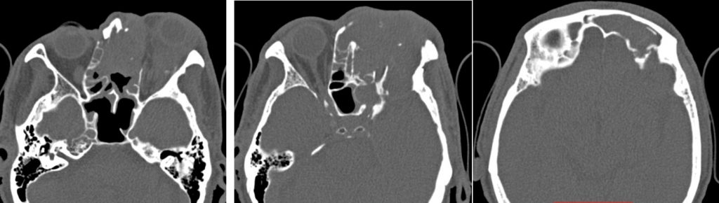
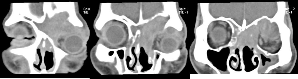
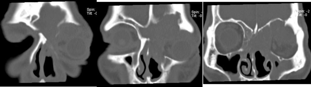
CT scan findings:
- Extensive soft tissue mass lesion in the left nasal cavity extending into the ethmoidal and frontal sinus, and also into left intraorbital cavity
- There is no obvious intracranial extension
- No calcification within the mass lesion
- It showed homogenous enhancement
- Extensive erosion of the adjacent bones are seen
- Mass effect causing proptosis of left eye
Diagnosis: Inverted papilloma (HPE proven)
Discussion:
- Inverted papilloma is a benign epithelial neoplasm that arises within the nasal vault and less commonly, in the paranasal sinuses.
- Inverted papillomas account for approximately 0.5-4.0% of all nasal tumors
- It is most frequently seen in patients 40-60 years of age
- There is slight male predilection
- The tumor is characterized by a high recurrence rate, associated epithelial malignant tumors (5-8%), and bone destruction.
- Malignant transformation occurs in about 10% of cases
- The CT appearance of inverted papilloma is variable and non-specific. Nonetheless, inverted papilloma is the most likely diagnosis when a unilateral mass in the nasal vault, producing benign bony changes, extends centrifugally into the maxillary and ethmoidal sinuses and through the nasal choana into the nasopharynx in an elderly patient with chronic nasal obstruction.
Recent Comments