
Clinical:
- A 37 years old lady
- Complaints of vague pain at suprapubic region
- Normal menstrual cycle
- No fever, no constitutional symptoms
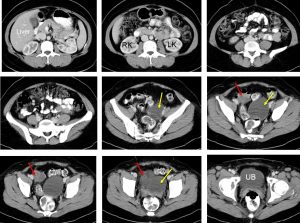
CT scan findings:
- There are two cystic lesions seen in the pelvic region
- The larger lesion at left side (yellow arrows) measuring about 6×7 cm, mainly cystic with no soft tissue component
- The smaller lesion at right side (red arrows) measuring 3×4 cm shows higher density. Presence of septa within it.
- No calcification or fat component within both lesions
- No ascites
Intra-operative findings:
- Adhesion between omentum and bowels with right and left ovarian tumour
- Left ovarian teratoma measuring about 8x8x5 cm, with cystic lesion ruptured during manipulation
- Right ovary with small endometriotic cyst measuring 3x2x2 cm also ruptured during manipulation
HPE findings:
- Macroscopy: specimen labelled as left adnexal consist of a piece of greyish tissue firm loculated surface measuring 60x25x20 mm. A part of fallopian tube attached to the mass. A serial section shows multiple cystic lesions measuring 3 mm to 20 mm in diameter contain gelatinous material. Another specimen labelled as right adnexal consist of a piece of ovarian tissue measuring 45x30x15 mm.
- Microscopy: section of the left ovarian tissue containing benign follicular cysts. Part of the ovarian stroma is congested and hemorrhagic. Section of the right ovary shows corpus luteum with haemorrhage in its lumen and surrounding stroma. There are a few ovarian follicles, some are cystic. No evidence of malignancy seen.
- Diagnosis: Right ovary: corpus haemorrhagicum and left ovary: benign follicular cyst
Diagnosis: Benign follicular cyst of ovary and corpus haemorrhagicum
Discussion:
- During each menstrual cycle, a very well synchronized sequence of events takes place surrounding the maturation and release of an oocyte.
- The process begins with the development of a cystic swelling up to 2.8 cm in diameter, known as a graafian follicle. At midcycle, after rupture and release of a mature oocyte, this follicle becomes a corpus luteum, a 1.5- to 2-cm structure with a cystic center.
- If fertilization occurs, the corpus luteum initially enlarges and elaborates progesterone until the placenta takes over that function at about 12 weeks’ gestation.
- If no pregnancy occurs, the corpus luteum generally involutes, but in some cases, it can persist and develop into a functional ovarian cyst (also known as a follicular cyst or cystic follicle).
- Grossly, these cysts are rarely greater than 8 cm, have glistening membrane, thin-walled and can be unilocular or multilocular. It demonstrate clear or serous fluid contents with no solid component
- The corpus haemorrhagicum (bleeding corpus luteum) is a temporary structure formed immediately after ovulation from the ovarian follicle as it collapses and is filled with blood that quickly clots.
Recent Comments