
Clinical:
- A 2 years old girl
- Previously noted to have swelling at neck region
- Clinically there is soft (cystic) lesion at lateral neck on the right side
- Later presented with discharge from a small opening at same area of neck region
Imaging findings:
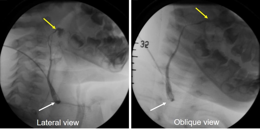
- Fistulogram performed; contrast injected via the opening at neck region (white arrows)
- The contrast is seen opacifying a tract along the lateral neck superiorly
- The second opening is seen at the region of palatine tonsil
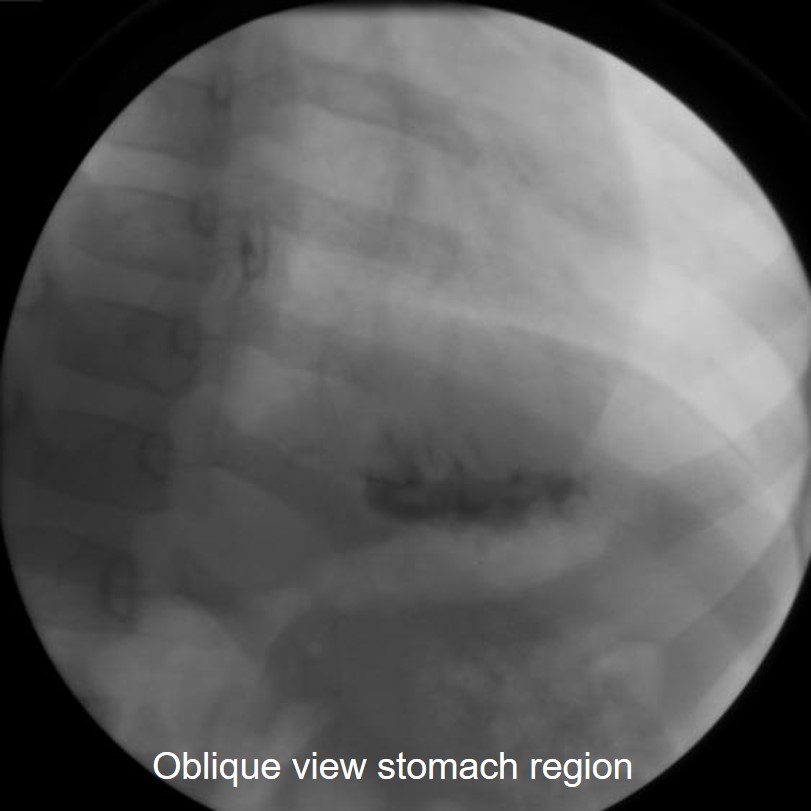
- Delayed image shows contrast in the stomach
Diagnosis: Second branchial cleft fistula.
Discussion:
- Second branchial cleft cysts makes up approximately 95% of all branchial anomalies.
- Although a congenital abnormality, second branchial cleft cyst tend to present in early adulthood (10-40 years of age) often after minor trauma or infection.
- However, second branchial cleft fistula usually present earlier.
- Fistulas extend from the skin surface anterior to the middle of the sternocleidomastoid muscle, pass between the ICA and ECA and eventually drain into the tonsillar fossa.
- Clinically the cysts are painless, fluctuant masses, situated at the lateral aspect of the neck, proximal to the anteromedial border of the sternocleidomastoid, at the mandibular junction.
- They gradually progress in size, and may become painful or tender with time if they are subsequently infected.
- On USG, it looks like an anechoic mass or chiefly hypoechoic cystic mass, with faint internal debris and posterior enhancement.
- On a CT scan, the cyst usually appears as a well-circumscribed, non-enhancing mass of homogeneous low attenuation.
-
On T1 MRI sequence, it shows variable signal depends on the protein content. It is usually hyperintense on T2 and shows no enhancement post contrast in non-complicated cases.
- Since there is a possibility of secondary infection, the preferred management strategy is surgical excision.