
Clinical:
- A 72 years old man
- Underlying HPT and hyperlipidaemia
- Presented with forgetfulness x 3/52.
- Associated with weakness at right UL and LL.
- Clinical examination shows receptive dysphasia. Power right UL and LL 4/5.
- Urgent plain CT scan brain requested to rule out CVA.
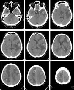
CT scan findings:
- Multiple round hyperdense lesions are seen at the grey-white matter junction in both cerebral hemispheres. The largest is seen at the right frontal region, measuring about 2.9(AP x 2.9(W) x 3.0(CC)cm.
- Some of these lesions demonstrate central hypodensity, likely suggestive of central necrosis.
- Marked perilesional white matter oedema with effacement of the adjacent sulci is seen. The body of the left lateral ventricle is slightly compressed.
- No midline shift. No hydrocephalus. Basal cisterns are not effaced.
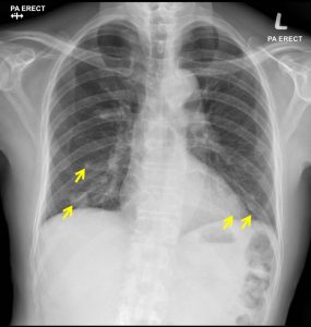
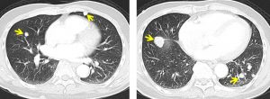
Radiographic findings:
- There are multiple well defined nodules seen in right and left lower zone
- The largest in right lower zone measured 2.2 x 2.0 cm.
- No consolidation. No pleural effusion or pneumothorax. No hilar enlargement seen. Heart size is within normal range.
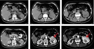
CT scan findings:
- There is a large heterogeneously enhancing lobulated mass seen occupying the upper and interpolar regions of the left kidney, measuring at 8.5 x 5.6 x 6.6cm (AP x W x CC). The mass vividly enhances post contrast with central hypodensities noted, possibly representing central necrotic cores. No calcification seen within the mass.
- The mass extends into the renal pelvis region. No hydronephrosis seen.
- The left renal vein is well opacified with no apparent filling defect seen within to suggest presence of thrombosis. The renal arteries appear unremarkable.
Diagnosis: Hyperdense cerebral metastasis from left renal carcinoma
Discussion:
- Cerebral metastases are estimated to account for approximately 25-50% of intracranial tumors in hospitalized patients.
- The term cerebraltechnically includes the cerebrum, the cerebellum and the brainstem.
- Hyperattenuating brain metastasis (on CT scan) can be caused by malignant melanoma, choriocarcinoma, colon cancer, renal cell cancer and thyroid cancer.
Progress of patient:
- Condition of patient deteriorated.
- Patient died one month after the presentation.