
Clinical:
- A 69 years old man with underlying DM, COAD
- Also had right nasal polyposis with sinusitis.
- Recently treated for right otitis externa with acute mastoiditis.
- After 2 weeks completed treatment patient complaint of hoarseness of voice
- Clinical examination shows patient having right vocal cord palsy.
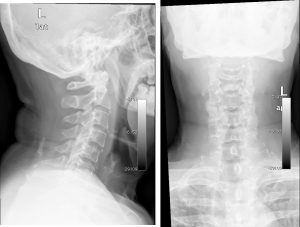
- CT scan of the neck region shows no significant findings.
- After 3 weeks start to have fever, right sided headache and progressive upper limb weakness.
CT scan findings:
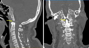
- Erosion at tip of dens seen.
- Otherwise no surrounding soft tissue mass
- No reduction in vertebral body heights.
MRI findings:
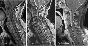
- MRI of neck shows abnormal signal intensity at C1 and C2 vertebrae
- There is diffuse enhancement on post contrast image at this region and also at the right mastoid tip (images not shown) with small abscess collection.
Progress of patient:
- Lumbar puncture done. CSF appearance clear, AFB stain negative, Indian Ink stain negative, polymorphs 8, lymphocyte 25/cmm, rbc 0, no growth after 48 hours, no MTB isolated after 6 weeks
- Mycobaterium tuberculosis complex PCR negative
- Biopsy of mastoid lesion showed a fragment of fibrous tissue densely infiltrated by lymphoplasma cells and neutrophils. Areas of granulation tissue formation and calcification are noted. No granuloma or malignancy seen. Ziehl Neelson stain for acid fast bacilli is negative.
Diagnosis: Cervical vertebrae osteomyelitis from mastoiditis.
Discussion:
- The incidence of spinal osteomyelitis that exclusively involved the
cervical spine ranged from 3 to 11%. - The mean age of the patients ranged from 57 to 60 years.
- A majority of patients in all series were men, with a range of 66 to 87%.
- Medical comorbidities or serious medical illnesses were identified in many of the patients (range 43–64%), but spontaneous cases of osteomyelitis were also reported.
- On MRI, spinal infections commonly demonstrate typical signal intensity on T1- and T2-weighted images and enhancement within the affected bone marrow after the administration of gadolinium-based contrast material.
Recent Comments