Clinical:
- A 2 years old boy
- History of trauma and right subdural bleed with cerebral oedema at the age of 5 months old.
- After that developed recurrent partial seizure.
- Also subsequently had delayed motor development
- MRI to see progress of head injury

CT scan findings:
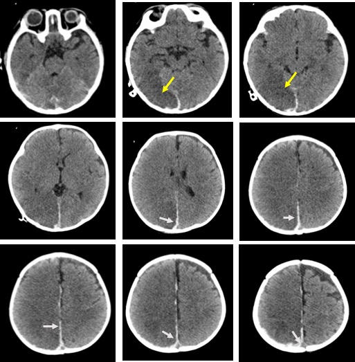
- Initial CT brain done one year ago
- There is hyperdensity along the falx on the right side in keeping with subdural bleed.
- Hypodensities seen in right cerebral hemisphere, predominantly in occipital and parietal lobes.
- Effacement of the cerebral sulci on the right side
- No significant midline shift. No uncal herniation. No hydrocephalus.
- No obvious skull vault fracture (bone window images not shown).


MRI findings:
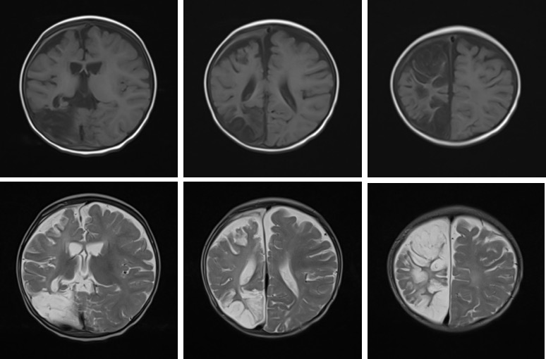
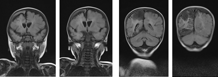
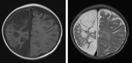
- There is abnormal signal intensity within the right frontal, parietal and occipital lobes as well as in the right lentiform nucleus, which is hypointense on T1and FLAIR, hyperintense on T2 and does not enhance post contrast, suggestive of encephalomalacic changes from previous insult.
- There is no significant midline shift to the right.
- No area of restricted diffusion.
- Chronic right subdural bleed.
Diagnosis: Encephalomalacia with chronic subdural hemorrhage.
Discussion:
- Encephalomalacia is a term given to describe loss of brain parenchyma with or without surrounding gliosis, as a late manifestation of injury
- Encephalomalacia occurs following insult, usually occurring aftercerebral ischaemia, cerebral infarction, hemorrhage, traumatic brain injury, surgery or other insults.
- By strict definition, gliosis is not synonymous with encephalomalacia. However, gliosis and encephalomalacia often coexist during the early and intermediate responses to injury, with gliosis waning with time.
- Encephalomalacia seen on CT scan as region of hypoattenuation, volume loss and can occur anywhere. owever, characteristic locations are anteroinferior frontal and temporal lobes.
- On MRI it follows CSF signal on all sequences, unlike gliosis which appears bright on T2 as well as FLAIR.
