
Clinical:
- A 69 years old lady
- History of recurrent right hypochondriac pain
- Current presentation with slight jaundice
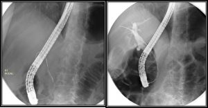
ERCP findings:
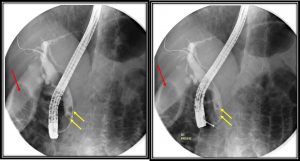
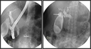
- There is filling defect seen within the gall bladder suggestive of calculus (red arrow).
- The cystic duct is dilated measuring about 9 mm (white arrow).
- The common duct is also dilated measuring about 13 mm.
- There are multiple filling defect at distal common duct in keeping with calculi (yellow arrows).
- The intrahepatic ducts are not dilated.
- The calculi within the common duct were removed during the procedure.
Diagnosis: Choledocholithiasis and cholelithiasis, endoscopic removal of choledocholithiasis.
Discussion:
- Choledocholithiasis is stone within the bile ducts (including the common hepatic duct/common bile duct).
- Choledocholithiasis is relatively common, seen in 6-12% of patients who undergo cholecystectomy.
- Stones within the bile duct may form either in-situ or pass from the gallbladder, and when recurrent tend to be pigment stones, and are thought to be associated with bacterial infection.
- Endoscopic retrograde cholangiopancreatography (ERCP) with sphincterotomy is the treatment of choice for choledocholithiasis.
- This procedure is associated with a complication rate of 5.8-24%.
Recent Comments