
Case contribution: Dr Radhiana Hassan
Clinical:
- A 55 years old lady
- Presented with right renal pain
- US KUB done shows a small right renal cyst with wall calcification
- For further assessment of the cyst
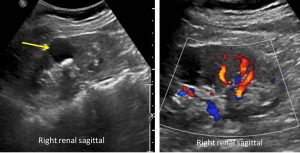
Ultrasound findings:
- There is cyst measuring 2.0 x 2.3 cm size in inter pole of right kidney.
- There is hyperechoic foci measuring 1.2 cm size within the lesion that lies in the periphery and at dependant region
- Posterior shadowing of the echogenic foci seen that could be calculus.
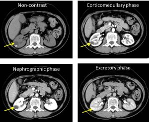
CT scan findings:
- A round hypodense cystic lesion is seen at right upper pole measuring 1.7 x 2.1 x 2.1 cm (AP x W x CC).
- The lesion has thin imperceptible wall and peripheral rim of wall calcification.
- A small calculus is seen within measuring 0.9 cm.
- On excretory phase the layering of contrast material is seen within likely due to communication with the upper pole major calyx.
- No other lesion in both kidneys. No hydronephrosis.
Diagnosis: Calyceal diverticulum in the right kidney.
Discussion:
- Calyceal diverticulum is outpouching from the renal calyx or renal pelvis into the renal cortex.
- It is also known as pyelocalyceal diverticulum
- It occurs equally in men and women.
- There is no predilection for the left or right side.
- The exact etiology is unknown but many favor congenital over acquired origin.
- It is classified as type I, those communicating with a minor calyx or an infundibulum and type II in those emanating from the renal pelvis or major calyx.
- Type I is the more common form calyx and is usually found in the upper pole
- Type II are larger, tends to be symptomatic and are located in the interpolar region of the kidney.
- Calyceal diverticulum is not seen on plain radiography.
- If calcifications are present, these may be visible as with other renal calculi.
- Ultrasound may reveal renal cyst but will not demonstrate its connection to the pelvicalyceal system.
- Features of echogenic sediment, irregular morphology of wall and material inside may suggest a diagnosis of diverticulum.
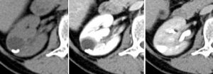
- On early phase CT scan, it appears as small round, low-attenuation areas adjacent to the calyces.
- Following contrast administration, layering of contrast in the dependant position is noted. The patency of the diverticulum is demonstrated by the gradual opacification on delayed scans
- A frequent complication associated with calyceal diverticulum is stones. Stone forms within the calyceal diverticula in up to 50% of cases.
- Other complication include infection, hemorrhage or transitional cell carcinoma arising from its uroepithelial lining.
- Management depends on symptoms
- Conservative management in asymptomatic patients.
- Intervention is considered if symptomatic.
Recent Comments