
Case contribution: Dr Radhiana Hassan
Clinical:
- A 43-year old lady
- No known medical illness
- Left breast lump felt for few months
- Sudden increase in size for the past 2 months
- No family history of breast cancer
- No fever, not painful, no nipple discharge
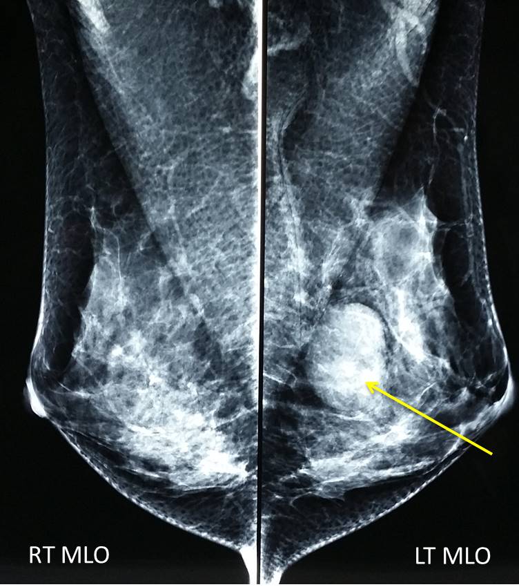
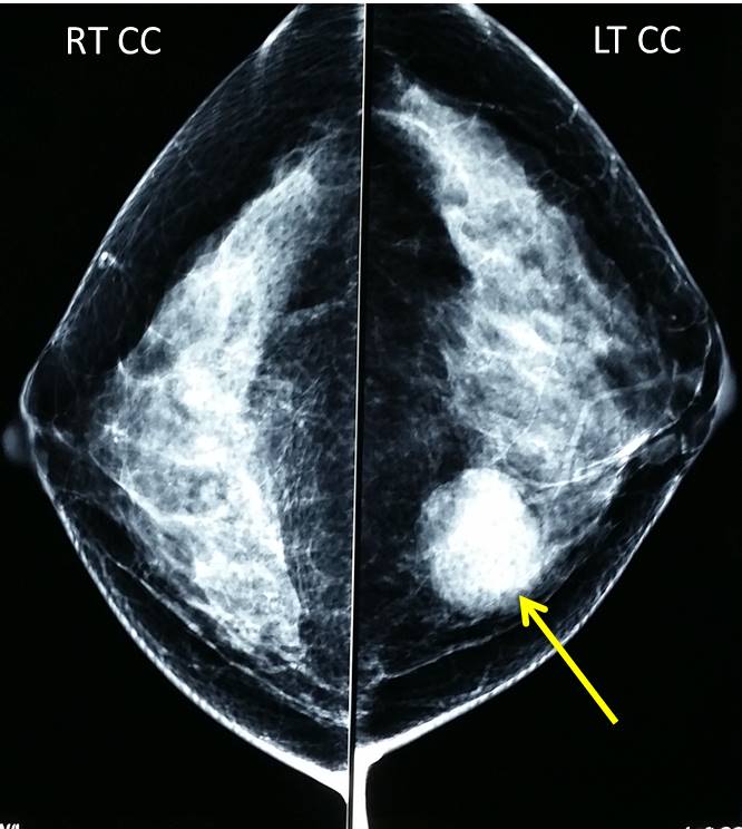
Mammogram findings:
- Bilaterally dense breasts
- A well-defined mass lesion is seen at left inner midquadrant (yellow arrows)
- The lesion measures 37×34 mm
- No stromal distortion or clustered microcalcifications
- No nipple retraction or skin thickening
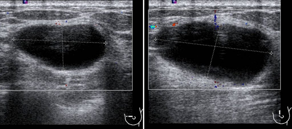
Ultrasound finding:
- A well defined hypoechoic lesion in Lt11H measuring 33x26x13 mm
- Posterior enhancement seen
- No internal echoes or debri
- No abnormal vascularity seen
Progress of patient:
- FNAC done at pathology lab
- Yellowish colour aspirate. Lesion totally collapsed after procedure. Hypocellular aspirates with occasional macrophages
- Impression: benign cysts with apocrine change
Diagnosis: Simple breast cyst
Discussion:
- Cystic lesions of the breast are common in women between 30-50 years old
- On mammography, a cystic lesion appears as a round, oval, or lobulated mass with circumscribed margins
- Mammogram alone cannot reliably diagnosed a cyst
- Ultrasound reliably differentiate cystic from solid lesions in most cases and permits further characterization of the mass; evaluation of the lesion’s shape, orientation, margin, boundary, internal echotexture, posterior acoustic features, surrounding tissue, calcifications, and vascularity.
- Simple cysts on ultrasound is seen as an anechoic, well circumscribed with a thin echogenic capsule, increased through-transmission, and thin edge shadows.
- Simple cyst should not increase in size in post-menopausal women.
- A simple cyst is classified as BI-RADS 2 and the subsequent follow up follows a screening protocol.
- Symptomatic large cysts may warrant aspiration. Simple cyst aspiration showing straw colored fluid can be discarded. Cytological analysis is usually not required, unless it contains bloody material.
- Post-aspiration ultrasound confirms the cyst has disappeared completely with no residual mass and will confirm hemostasis.
- Complications from aspiration are virtually unknown but include bleeding and theoretically infection.
- Aspiration of cysts can be safely performed without stopping aspirin therapy.
Reference:
- Hines et al. Cysts masses of the breast. AJR 2010: 194:W122-W133
- Kabbani et al. Simple breast cyst. Radiopedia at https://radiopaedia.org/articles/simple-breast-cyst-1
Recent Comments