
Case contribution: Dr Radhiana Hassan
Clinical:
- A 58 years old lady
- Autoimmune hemolytic anemia (AIHA) under follow up
- NO DM, no HPT
- Left PUJ stone, laser endopyelorotomy, laser lithotripsy and stenting done one month ago
- Discharged well
- Presented with left lumbar pain and fever for 2 days
- Hb: 6.4, TWBC 20.0
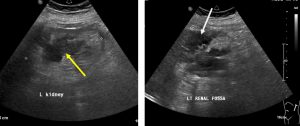
Ultrasound findings:
- A hypoechoic lesion in the left renal cortex (yellow arrow)
- Irregular outline with internal echoes
- A collection is seen outside the left renal (white arrow)
- No other significant finding
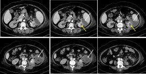
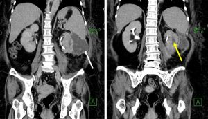
CT scan findings:
- The left kidney is shrunken with bipolar length (BPL) of 7.4cm.
- Multiple calculi are seen in the upper and lower pole of the left kidney.
- There are multi-loculated peripherally enhancing lesions within the left renal cortex. The largest loculation is seen within the midpole, peripherally, measuring about 1.6cm x 2.4cm x 3.8cm (APxWxCC).
- A larger peripherally enhancing collection is seen within the splenorenal region measuring about 7.0cm x 7.9cm x 8.3cm (APxWxCC). Multiple small air pockets noted.
- This collection is seen connected to the left renal collection at the anterolateral aspect of the left kidney.
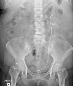
- Double – J stent is seen in situ.
Diagnosis: Left renal abscesses with perinephric collection
Discussion:
- Renal abscess is usually a sequalae of pyelonephritis
- On ultrasound, it appears as a well-defined hypoechoic area within the cortex or in the corticomedullary parenchyma. It demonstrates internal echoes within and an associated diffusely hypoechoic kidney due to acute pyelonephritis may be seen.
- Perinephric collection may also be seen.
- On CT scan it appears as a well-defined mass of low attenuation with a thick, irregular wall or pseudo capsule. Gas within a low attenuation/cystic mass strongly suggests abscess formation.
- Associated fascial and septal thickening is seen with obliteration of perinephric fat.
Recent Comments